Review
. 2017 Jun;1864(6):900-906.
doi: 10.1016/j.bbamcr.2016.11.028. Epub 2016 Nov 30.

The functions of store-operated calcium channels

Affiliations
Review

The functions of store-operated calcium channels

James W Putney et al. Biochim Biophys Acta Mol Cell Res. 2017 Jun.

Abstract

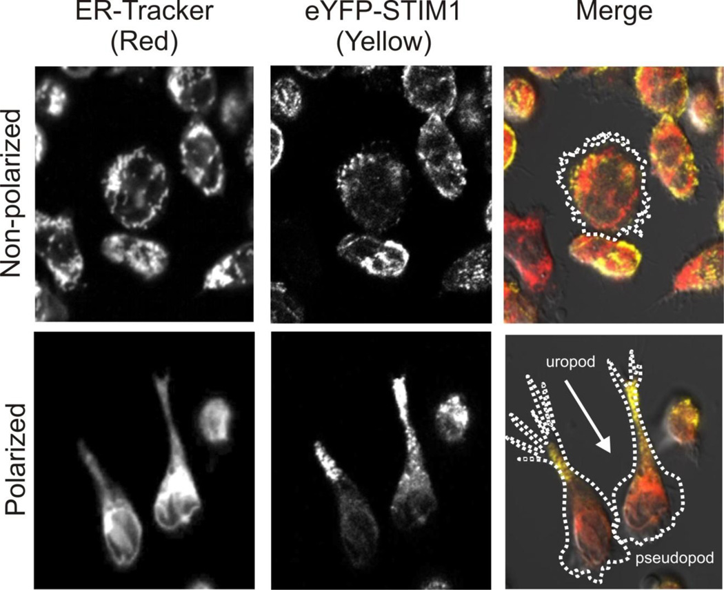
Store-operated calcium channels provide calcium signals to the cytoplasm of a wide variety of cell types. The basic components of this signaling mechanism include a mechanism for discharging Ca2+ stores (commonly but not exclusively phospholipase C and inositol 1,4,5-trisphosphate), a sensor in the endoplasmic reticulum that also serves as an activator of the plasma membrane channel (STIM1 and STIM2), and the store-operated channel (Orai1, 2 or 3). The advent of mice genetically altered to reduce store-operated calcium entry globally or in specific cell types has provided important tools to understand the functions of these widely encountered channels in specific and clinically important physiological systems. This review briefly discusses the history and cellular properties of store-operated calcium channels, and summarizes selected studies of their physiological functions in specific physiological or pathological contexts. This article is part of a Special Issue entitled: ECS Meeting edited by Claus Heizmann, Joachim Krebs and Jacques Haiech.

Keywords: Calcium signaling; Exocrine glands; Keratinocytes; Mouse models; Neutrophils; Store-operated calcium channels.


Conflict of interest statement

The authors declare there are no conflicts of interest.

Figures

Figure 1
Figure 1. STIM1 distributes to the rear of polarized HL-60 neutrophils
Neutrophil-like HL-60 cells stably expressing eYFP-STIM1 [99] were stained with the endoplasmic reticulum labeling reagent, ER-Tracker Red (Molecular Probes). Cells were plated on coverslips covered with fibronectin (polarized cells) or fibronectin plus BSA (nonpolarized cells). In polarized cells, STIM1 clearly distributes to the rear (uropod), and to a significantly greater extent than the endoplasmic reticulum.
Figure 2
Figure 2. Wound healing in wild-type and in mice with epidermal-specific knockout of STIM1
Methods: Mice were crossed with KRT14-cre mice (Jackson laboratories). Isolation of keratinocytes was carried out as previously demonstrated [117]. Keratinocytes were cultured in CnT-07 (Zen-bio) according to manufacturer’s instruction. Calcium imaging of keratinocytes were carried out as previously reported [103]. For superficial wounding of the epidermis, mice were subjected to a tape-stripping assay. 7–9 week old female mice were anesthetized with isoflurane. Mice backs were shaved and depilated by Nair cream. Depilated back skins were tape-stripped 20 times with Scotch tape (18 mm width). Lesions were rubbed with Vaseline to be moisturized. At the end of time-courses, lesioned skin was harvested for histological analysis. A: Epidermal specific knockout of STIM1 (STIM1-epKO) results in almost complete disappearance of STIM1 protein. B: Keratinocytes from STIM1-KO mice lack Ca2+ entry in response to thapsigargin or to elevated Ca2+. C: Left panel shows an example of the wound area produced by tape stripping. Right panel shows examples of injured area in a wild type (WT) and knock-out (KO) mouse at 24 hours and 120 hours. D: Histological examination of skin at 120 hours shows extensive hyperkeratinization of skin of wild type mouse not the knockout (STIM1-epKO) mouse. E. Summary data show that skin from knockout (STIM1-epKO) mice has a slightly decreased hyperplasia score and a significantly diminished mononuclear cell infiltrate and hyperkeratosis score (P<0.05).

References

    1. Putney JW. Recent breakthroughs in the molecular mechanism of capacitative calcium entry (with thoughts on how we got here) Cell Calcium. 2007;42:103–110. - PMC - PubMed
    1. Putney JW. Muscarinic, alpha-adrenergic and peptide receptors regulate the same calcium influx sites in the parotid gland. J. Physiol. (Lond.) 1977;268:139–149. - PMC - PubMed
    1. Parod RJ, Putney JW. The role of calcium in the receptor mediated control of potassium permeability in the rat lacrimal gland. J. Physiol. (Lond. ) 1978;281:371–381. - PMC - PubMed
    1. Casteels R, Droogmans G. Exchange characteristics of the noradrenaline-sensitive calcium store in vascular smooth muscle cells of rabbit ear artery. J. Physiol. (Lond. ) 1981;317:263–279. - PMC - PubMed
    1. Putney JW. A model for receptor-regulated calcium entry. Cell Calcium. 1986;7:1–12. - PubMed

Publication types