Review
. 2016 Dec 2;2016(1):217-225.
doi: 10.1182/asheducation-2016.1.217.

Atypical hemolytic uremic syndrome

Affiliations
Review

Atypical hemolytic uremic syndrome

Vahid Afshar-Kharghan. Hematology Am Soc Hematol Educ Program. .

Abstract

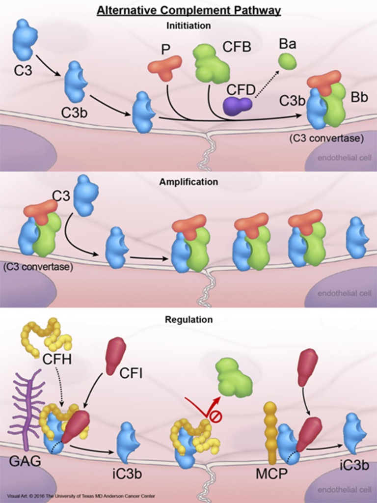
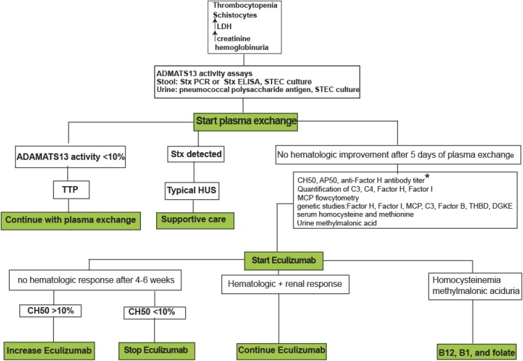
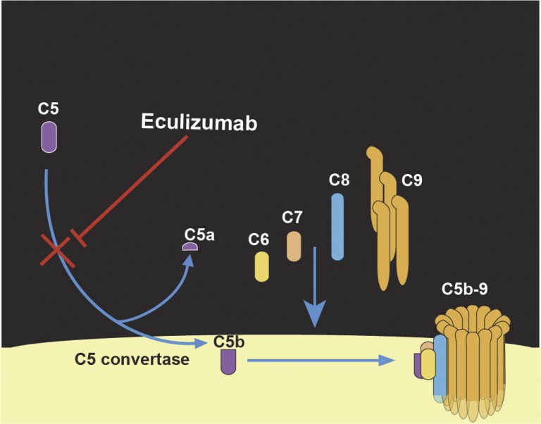
Atypical hemolytic uremic syndrome (aHUS) is a thrombotic microangiopathy (TMA) that affects multiple organs and the kidneys in particular. aHUS can be sporadic or familial and is most commonly caused by dysregulation of the alternative complement pathway. The initial attack of aHUS can occur at any age, and is associated with a high rate of progression to end stage renal disease. Many aHUS patients relapse in the native or transplanted kidneys, and require close monitoring and long-term management. Availability of anticomplement therapy has revolutionized the management of aHUS, and can change the natural course of aHUS by inducing hematologic remission, improving or stabilizing kidney functions, and preventing graft failure. As a result, it is important to succeed in the challenging task of differentiating aHUS from other TMAs and initiate adequate treatment early during the course of disease. Considering the high cost of currently available anticomplement therapy, it is important also from a financial point of view to accurately diagnose aHUS early during the course of disease and determine the necessary length of therapy. This highlights the need for development of precise complement functional and genetic studies with rapid turnaround time.


Conflict of interest statement

Conflict-of-interest disclosure: V.A.-K. is on the Board of Directors or on an advisory committee for Alexion.

Figures

Figure 1.
Figure 1.
Activation and regulation of the alternative complement pathway. A small amount of plasma C3 is continuously converted to C3b that is highly active and binds to cell surface via forming thiol bonds. Under normal conditions, C3b is rapidly degraded in plasma by FI and FH (FI cofactor), or on the cell surface by FI and glycosoaminoglycan-bound FH, membrane protein CD46 (or MCP), or CD35. Degradation of C3b to an inactive product (iC3b) on the cell membrane by membrane-bound FH, MCP, or CD35 requires FI, and is known as cofactor activity. IC3b cannot participate in any further complement activation. If not inactivated, C3b is able to bind to an activation product of FB (Bb) produced by FD-mediated cleavage of FB, and generate C3bBb (C3 convertase). C3 convertase is the engine of the complement pathways and deposit additional C3b molecules on membrane, which in turn amplify complement activation. C3 convertase is an important target for complement regulatory proteins. FH prevents the formation of C3 convertase and dissociate C3 convertase by competing with FBb for binding to C3b. This negative regulatory activity of FH is known as decay accelerating activity. If C3bBb remains intact, the complex is stabilized by FP (properdin) and binds to additional C3b to generate C3bBbC3b, which is also known as C5 convertase. C5 convertase activates C5 to generate C5b, which in turn binds to C6, C7, and C8, forming the C5b-8 complex that stably inserts into the lipid bilayer of the cell. Next, the C5b-8 complex binds and polymerizes multiple molecules of C9 (C5b-9n), forming cytolytic MAC of complement on the cell surface. CFB, factor B; CFD, factor D; CFH, factor H; CFI, factor I; GAG, glycosoaminoglycans; MAC, membrane attack complex; P, properdin. Reprinted from The University of Texas MD Anderson Cancer Center with permission.
Figure 2.
Figure 2.
Diagnostic and therapeutic algorithm in TMAs. Role of eculizumab in the treatment of secondary aHUS (cancer-, chemotherapy-, solid-organ transplant- or HSCT-, pregnancy-, or autoimmune disorder-associated) have not been established and is out of the scope of this study. *In patients with anti-FH antibody, immunosuppressive reagents (steroid, rituximab, cyclophosphamide, and azathioprine) should be added to eculizumab. ELISA, enzyme-linked immunosorbent assay; LDH, lactate dehydrogenase; PCR, polymerase chain reaction.
Figure 3.
Figure 3.
Mechanism-of-action of eculizumab, a humanized monoclonal antibody that binds to C5 with a high affinity and inhibits cleavage of C5 by C5 convertase, and prevents generation of C5a and formation of C5b-9 MAC.

References

    1. George JN, Nester CM. Syndromes of thrombotic microangiopathy. N Engl J Med. 2014;371(7):654-666. - PubMed
    1. Moake JL. Thrombotic microangiopathies. N Engl J Med. 2002;347(8):589-600. - PubMed
    1. Tarr PI, Gordon CA, Chandler WL. Shiga-toxin-producing Escherichia coli and haemolytic uraemic syndrome. Lancet. 2005;365(9464):1073-1086. - PubMed
    1. Schwartz J, Winters JL, Padmanabhan A, et al. . Guidelines on the use of therapeutic apheresis in clinical practice-evidence-based approach from the Writing Committee of the American Society for Apheresis: the sixth special issue. J Clin Apher. 2013;28(3):145-284. - PubMed
    1. Loirat C, Frémeaux-Bacchi V. Atypical hemolytic uremic syndrome. Orphanet J Rare Dis. 2011;6:60. - PMC - PubMed

MeSH terms

Substances