

What can the microstructure of stones tell us?
- PMID: 27913855
- PMCID: PMC5253090
- DOI: 10.1007/s00240-016-0944-z
What can the microstructure of stones tell us?
Abstract
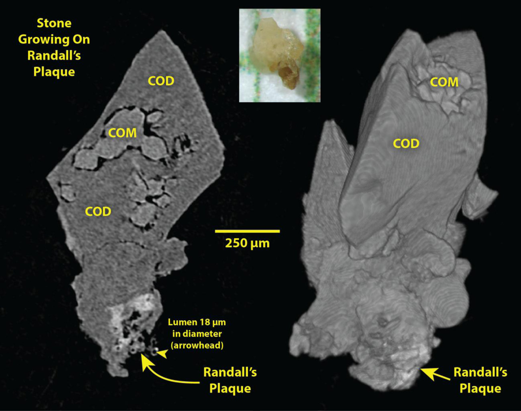
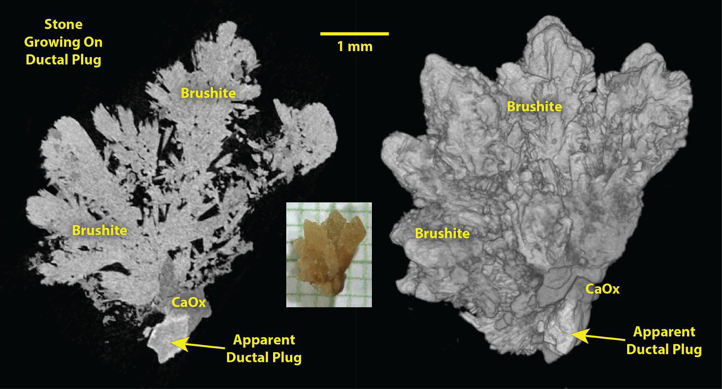
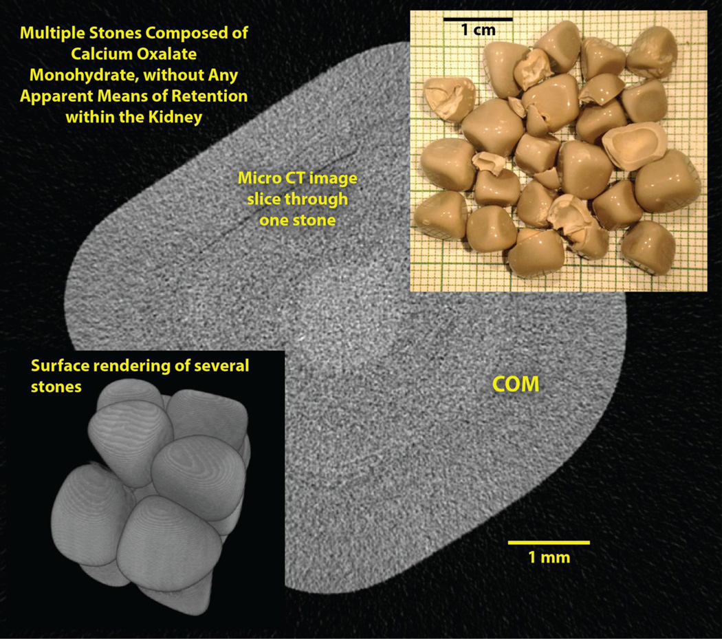
How stones are retained within the kidney while small in size is still not fully understood. In this paper, we show two examples of how stones are retained during early growth: one is growth on Randall's (interstitial) plaque, and the other is growth on mineral that has formed as a luminal plug in a terminal collecting duct. These two mechanisms of stone retention during early growth have distinctive morphologic features that can be seen by methods that show the microscopic structure of the stones. Stones growing on Randall's plaque display an apatite region that is typically not large in size (<0.5 mm across) but which usually shows luminal spaces, which are signs of its origin in the connective tissue of the papilla. Stones growing on ductal plugs also show attachment to a piece of apatite, but the apatite regions are typically larger (often >1 mm long and >0.5 mm wide), and they are solid, without spaces running through them. We propose that knowing the mechanisms of stone retention during early stone formation could allow for better treatment of stone diseases.
Keywords: Imaging; Nephrolithiasis.
Conflict of interest statement
none
Figures
References
-
- Robertson WG, Peacock M, Nordin BE. Activity products in stone-forming and non-stone-forming urine. Clinical Science. 1968;34:579–594. - PubMed
-
- Daudon M, Jungers P. Clinical value of crystalluria and quantitative morphoconstitutional analysis of urinary calculi. Nephron Physiol. 2004;98(2):31–36. - PubMed
-
- Evan AP, Lingeman JE, Coe FL, Shao YZ, Parks JH, Bledsoe SB, Worcester EM, Sommer AJ, Paterson RF, Kuo RL, Grynpas M. Apatite deposits in collecting duct lumens produce epithelial cell injury and interstitial inflammation in patients forming brushite renal stones. J Urol. 2004;171(4 suppl. S):299. (abstract)
Publication types
MeSH terms
Substances
Grants and funding
LinkOut - more resources
Full Text Sources
Other Literature Sources
