. 2017 Jan 15:228:141-146.
doi: 10.1016/j.virusres.2016.11.031. Epub 2016 Nov 30.

Detection of human immunodeficiency virus RNAs in living cells using Spinach RNA aptamers

Affiliations

Detection of human immunodeficiency virus RNAs in living cells using Spinach RNA aptamers

Brandon D Burch et al. Virus Res. .

Abstract

Many techniques currently used to measure HIV RNA production in cells suffer from limitations that include high background signal or the potential to destroy cellular context. Fluorophore-binding RNA aptamers offer the potential for visualizing RNAs directly in living cells with minimal cellular perturbation. We inserted a sequence encoding a fluorophore-binding RNA aptamer, known as Spinach, into the HIV genome such that predicted RNA secondary structures in both Spinach and HIV were preserved. Chimeric HIV-Spinach RNAs were functionally validated in vitro by testing their ability to enhance the fluorescence of a conditional fluorophore (DFHBI), which specifically binds Spinach. Fluorescence microscopy and PCR were used to verify expression of HIV-Spinach RNAs in human cells. HIV-1 gag RNA production and fluorescence were measured by qPCR and fluorometry, respectively. HIV-Spinach RNAs were fluorometrically detectable in vitro and were transcribed in human cell lines and primary cells, with both spliced and unspliced species detected by PCR. HIV-Spinach RNAs were visible by fluorescence microscopy in living cells, although signal was reproducibly weak. Cells expressing HIV-Spinach RNAs were capable of producing fluorometrically detectable virions, although detection of single viral particles was not possible. In summary, we have investigated a novel method for detecting HIV RNAs in living cells using the Spinach RNA aptamer. Despite the limitations of the present aptamer/fluorophore combination, this is the first application of this technology to an infectious disease and provides a foundation for future research into improved methods for studying HIV expression.

Keywords: Aptamers; HIV-RNA; Spinach.


Figures

Figure 1
Figure 1. HIV RNAs containing Spinach are detectable in vitro
(A) Insertion sites for Spinach1/2 within HIV. (B) Images of colorless microcentrifuge tubes containing selection buffer + DMSO/DFHBI +/- the indicated RNAs, exposed to 365 nm light. (C) Emission spectra generated by TAR-Spinach1MATGmut or TAR RNAs + DFHBI, or DFHBI alone, upon exposure to 425 nm light. Emission is displayed in arbitrary units with peak emission of TAR-Spinach1MATGmut set to 100. (D) Fluorometry values, normalized to Spinach1MATGmut, of the indicated RNAs + DFHBI, or DMSO/DFHBI alone. (E) Fluorometry values, normalized to Spinach1MATGmut (left panel) or Spinach2ATGmut (right panel) for the indicated RNAs + DFHBI. HIV-Spinach RNAs contain 600-1000 nucleotides of HIV sequence. (F) in vitro RNA folding (method of Strack et al., 2013), normalized to tRNA-Spinach2, for the indicated RNAs at 25°C.
Figure 2
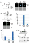
Figure 2. HIV-Spinach RNAs are detectable in cells and give rise to fluorometrically-detectable virions
(A) Gel of PCR product amplified from cDNA synthesized from total RNA extracted 72 h post-transfection from HeLa cells expressing the indicated constructs (left) and fluorescence spectroscopy images of a subset of these cells (right). (B) Gel of PCR product amplified from cDNA synthesized from total RNA extracted 72 h post-transfection from 293T cells expressing the indicated constructs (top) and fluorescence spectroscopy images of a subset of these cells (bottom). (C) HIV-1 Gag RNA copy number was detected by quantitative PCR off of cDNA produced from 500 ng total RNA from untransfected 293T cells (no HIV) or 293T cells transfected with DHIV3-TAR-Spin1MATGmut or DHIV3-TAR-Spin2ATGmut (from a different, but related experiment to the panels in (B)) (D) Fluorometry values, in arbitrary units, of HIV virions containing a Spinach1MATGmut aptamer within the 5’ TAR region + DMSO/DFHBI. DFBHI alone acts as a negative control. (E) Gel of PCR product amplified from genomic DNA isolated from CD4+ T cells infected with DHIV3-TAR-Spinach1MATGmut virions (top) and gel of PCR product amplified from cDNA synthesized from total RNA extracted from these cells (bottom). Note: PCR primer sequences are provided in Table S1. A schematic of the target locations for total, unspliced, and multiply spliced primer pairs is provided in Figure S2. A summary of mean qPCR and ELISA results for all tested constructs, including controls, is provided in Table S2.

References

    1. Bertrand E, Chartrand P, Schaefer M, Shenoy SM, Singer RH, Long RM. Localization of ASH1 mRNA particles in living yeast. Molecular cell. 1998;2(4):437–445. - PubMed
    1. Bosque A, Planelles V. Induction of HIV-1 latency and reactivation in primary memory CD4+ T cells. Blood. 2009;113(1):58–65. - PMC - PubMed
    1. Chable-Bessia C, Meziane O, Latreille D, Triboulet R, Zamborlini A, Wagschal A, Jacquet JM, Reynes J, Levy Y, Saib A, Bennasser Y, Benkirane M. Suppression of HIV-1 replication by microRNA effectors. Retrovirology. 2009;6:26. - PMC - PubMed
    1. Chizzolini F, Forlin M, Cecchi D, Mansy SS. Gene position more strongly influences cell-free protein expression from operons than T7 transcriptional promoter strength. ACS synthetic biology. 2014;3(6):363–371. - PubMed
    1. Filonov GS, Jaffrey SR. RNA Imaging with Dimeric Broccoli in Live Bacterial and Mammalian Cells. Curr Protoc Chem Biol. 2016;8(1):1–28. - PMC - PubMed

Publication types