Review
. 2017 Jun:107:216-227.
doi: 10.1016/j.freeradbiomed.2016.11.050. Epub 2016 Nov 30.

DNA damage related crosstalk between the nucleus and mitochondria

Affiliations
Review

DNA damage related crosstalk between the nucleus and mitochondria

Mohammad Saki et al. Free Radic Biol Med. 2017 Jun.

Abstract

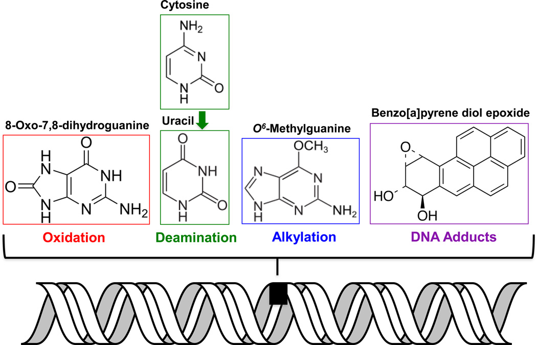
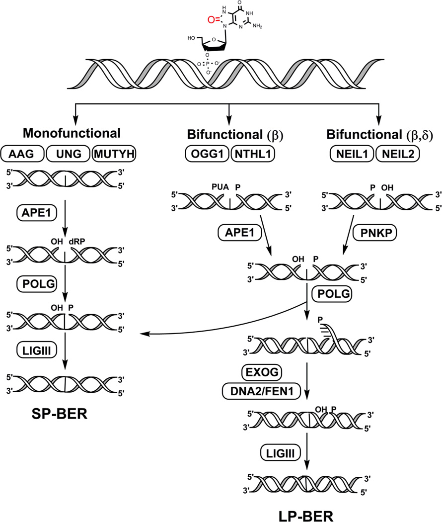
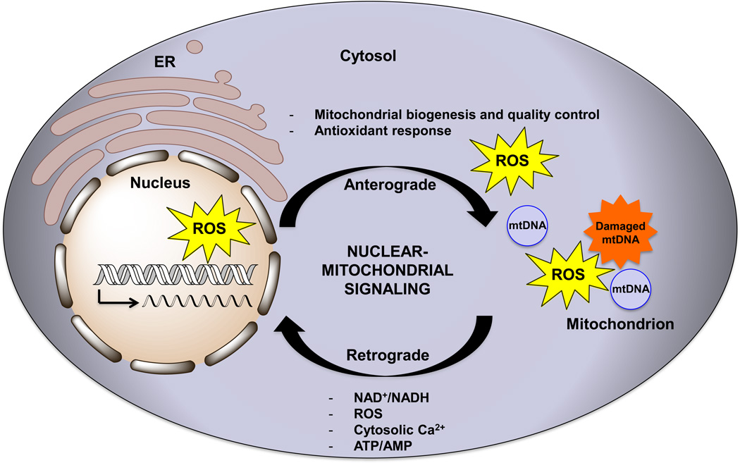
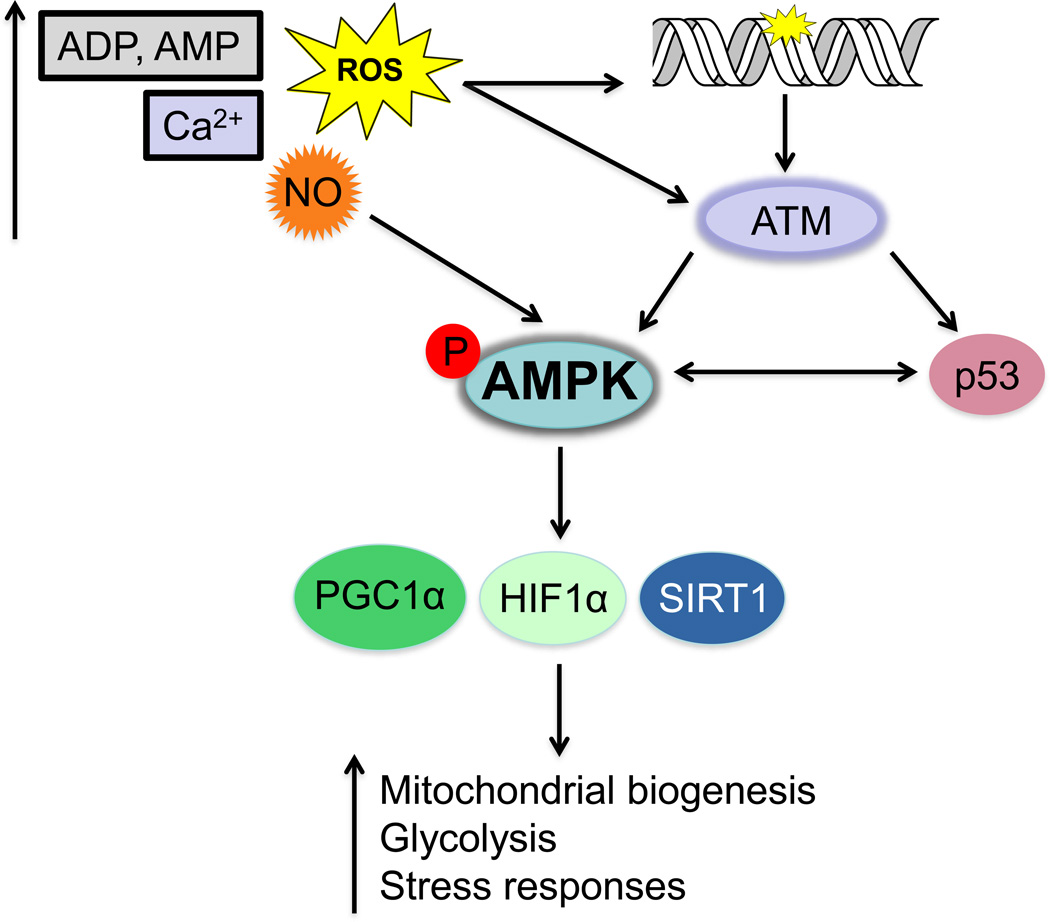
The electron transport chain is the primary pathway by which a cell generates energy in the form of ATP. Byproducts of this process produce reactive oxygen species that can cause damage to mitochondrial DNA. If not properly repaired, the accumulation of DNA damage can lead to mitochondrial dysfunction linked to several human disorders including neurodegenerative diseases and cancer. Mitochondria are able to combat oxidative DNA damage via repair mechanisms that are analogous to those found in the nucleus. Of the repair pathways currently reported in the mitochondria, the base excision repair pathway is the most comprehensively described. Proteins that are involved with the maintenance of mtDNA are encoded by nuclear genes and translocate to the mitochondria making signaling between the nucleus and mitochondria imperative. In this review, we discuss the current understanding of mitochondrial DNA repair mechanisms and also highlight the sensors and signaling pathways that mediate crosstalk between the nucleus and mitochondria in the event of mitochondrial stress.

Keywords: Mito-nuclear signaling; Mitochondrial DNA damage; Mitochondrial DNA repair; Mitochondrial dysfunction; Oxidative phosphorylation; REDOX signaling; Reactive oxygen species.


Conflict of interest statement

Conflict of Interest

None

Figures

Figure 1
Figure 1. Types of DNA lesions found in the mitochondria
Damage to mtDNA can occur in the form of alkylation, oxidation, spontaneous deamination, and bulky adducts. Examples of lesions from each of these categories are shown.
Figure 2
Figure 2. Overview of base excision repair in the mitochondria
This figure displays the step-wise repair of DNA base damage via the BER pathway by enzymes identified in the mitochondria. Initially, the lesion is recognized by either mono or bifunctional DNA glycosylases depending on the type of damage. Of the eleven known mammalian DNA glycosylases, only seven have been identified in the mitochondria. These include: the monofunctional glycosylases AAG (alkyladenine DNA glycosylase), UNG (uracil N-glycosyalse), and MUTYH (MutY glycosylase homolog) as well as the bifunctional glycosylases OGG1 (8-oxoG DNA glycosylase 1), NTHL1 (Nth-Like 1), NEIL1 (Nei-like 1), and NEIL2 (Nei-like 2). The resulting AP sites are further processed either by APE1 in case of the monofunctional, OGG1, and NTHL1 glycosylases or by PNKP that processes the ends after the NEIL enzymes, thereby leaving suitable ends for gap-filling by POLG. Ligase III then seals the DNA nick and completes the process in SP-BER. In the long-patch (or LP, >2 or more nt) repair pathway, a 2–6 nt flap is generated by POLG that is further processed by DNA2/FEN1. Alternatively, EXOG may function as the major 5’ flap-processing enzyme in the mitochondria in both SP- and LP-BER. The final ligation step is carried out by LIGIII.
Figure 3
Figure 3. Signaling between the nucleus and mitochondria
Crosstalk between the nucleus and mitochondria occurs not only to signal oxidative stress, DNA damage, and mitochondrial dysfunction but also occurs during normal cellular metabolism. The flow of information from the nucleus to the mitochondria (termed anterograde signaling) involves the transcription and translocation of genes involved with mitochondrial biogenesis. Anterograde regulation also includes responses to stressors that trigger an antioxidant response by regulating the expression of genes involved with Ca2+ metabolism and glycolysis. Mitochondria can signal to the nucleus (in a process called retrograde signaling) in times of stress via signals such as changes in the levels of NAD+/NADH, ROS, cytosolic Ca2+, and ATP/AMP as well as changes in membrane potential. ER stands for endoplasmic reticulum. The arrow within the nucleus signifies the transcriptional activation of nuclear genes either during normal conditions, or upon stress induced signaling.
Figure 4
Figure 4. Schematic of AMP-activated kinase (AMPK) signaling triggered by changes in cellular metabolites and DNA damage
AMPK is activated upon increased levels of AMP/ADP, ROS, and cytosolic calcium. Activation of AMPK can be triggered by the ataxia telangiectasia mutated (ATM) kinase that is activated upon oxidative stress or DNA damage. ATM triggers a DNA damage response that activates other factors such as p53 that can also directly activate AMPK. The activation of AMPK elicits downstream responses regulated by proteins including PGC-1α, SIRT1 and HIF-1α. These responses manifest in an upregulation of mitochondrial processes such as glycolysis, fatty acid oxidation, and responses to hypoxia.

References

    1. Pederson T. The nucleus introduced. Cold Spring Harb Perspect Biol. 2011;3(5) - PMC - PubMed
    1. Ernster L, Schatz G. Mitochondria: a historical review. J Cell Biol. 1981;91(3 Pt 2):227s–255s. - PMC - PubMed
    1. Gray MW. Mitochondrial evolution. Cold Spring Harb Perspect Biol. 2012;4(9):a011403. - PMC - PubMed
    1. Andersson SG, Karlberg O, Canback B, Kurland CG. On the origin of mitochondria: a genomics perspective. Philos Trans R Soc Lond B Biol Sci. 2003;358(1429):165–177. discussion 177-9. - PMC - PubMed
    1. Kowaltowski AJ. Alternative mitochondrial functions in cell physiopathology: beyond ATP production. Braz J Med Biol Res. 2000;33(2):241–250. - PubMed

Publication types

Substances