Review
. 2017 Jan 20;429(2):177-191.
doi: 10.1016/j.jmb.2016.11.024. Epub 2016 Dec 1.

Deciphering, Communicating, and Engineering the CRISPR PAM

Affiliations
Review

Deciphering, Communicating, and Engineering the CRISPR PAM

Ryan T Leenay et al. J Mol Biol. .

Abstract

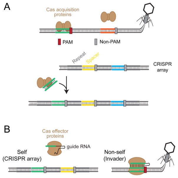
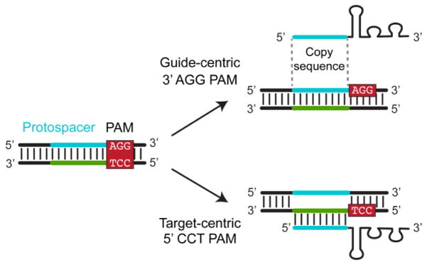
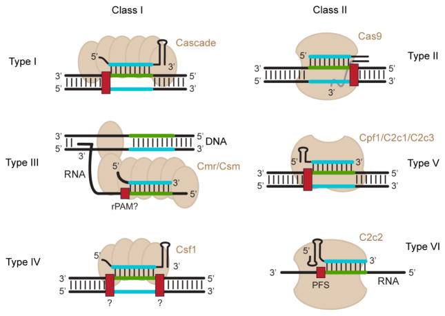
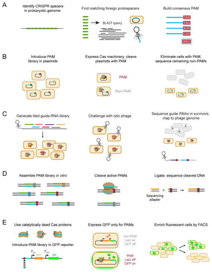
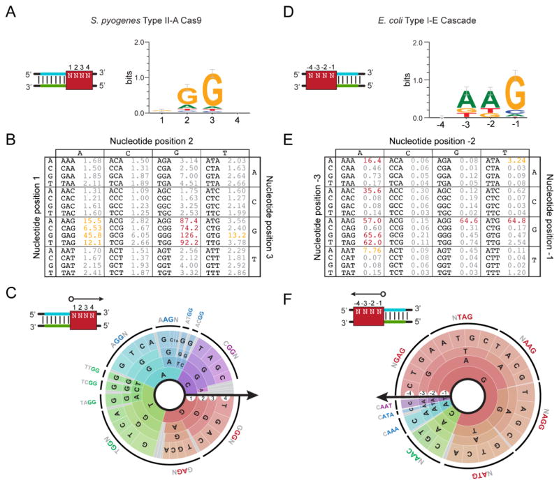
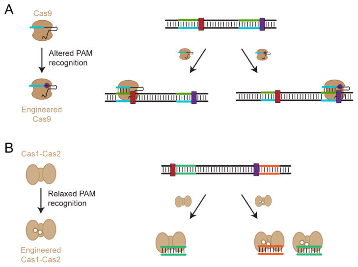
Clustered regularly interspaced short palindromic repeat (CRISPR) loci and their flanking CRISPR-associated (cas) genes make up RNA-guided, adaptive immune systems in prokaryotes whose effector proteins have become powerful tools for basic research and biotechnology. While the Cas effector proteins are remarkably diverse, they commonly rely on protospacer-adjacent motifs (PAMs) as the first step in target recognition. PAM sequences are known to vary considerably between systems and have proven to be difficult to predict, spurring the need for new tools to rapidly identify and communicate these sequences. Recent advances have also shown that Cas proteins can be engineered to alter PAM recognition, opening new opportunities to develop CRISPR-based tools with enhanced targeting capabilities. In this review, we discuss the properties of the CRISPR PAM and the emerging tools for determining, visualizing, and engineering PAM recognition. We also propose a standard means of orienting the PAM to simplify how its location and sequence are communicated.

Keywords: CRISPR–Cas systems; Cas9; Cpf1; PFS; rPAM.


Figures

Figure 1
Figure 1
Function of the CRISPR PAM. CRISPR-Cas systems naturally utilize PAMs to discriminate between self and non-self. The spacer portion of the CRISPR RNA is perfectly complementary to both its own CRISPR array (self) and the protospacer within the foreign invader’s genetic material (non-self). The systems differentiate between these two through the flanking PAM that is absent within the CRISPR array (gray) and present within the invader’s genetic material (red). Because the PAM is essential for target recognition, the CRISPR-Cas systems will target the invader’s genetic material but not the CRISPR array.
Figure 2
Figure 2
Two orientations for reporting PAM sequences. The PAM sequence is located within double-stranded DNA, where either strand of the PAM can be reported along with its location relative to the protospacer. Under the target-centric orientation, the PAM is reported from the strand that base pairs with the guide RNA. Under the guide-centric orientation, the PAM is reported from the strand that matches the guide RNA and is used for guide-RNA design.
Figure 3
Figure 3
PAM orientation and targets for different types of CRISPR-Cas systems. CRISPR-Cas systems are subdivided into two classes and six types. Representative illustrations of the effector proteins for each type are shown. The dsDNA orientation has been flipped from Figure 2 to accommodate the guide-centric PAM orientation. Note that the effector proteins for the V-B and V-C subtypes require a tracrRNA similar to Type II systems. For Type III and VI systems, the crRNA binds target RNAs. Type III systems cleave transcribed dsDNA due to its proximity to the RNA target [40]. Note that the mechanism by which Type III and VI systems recognize their nucleic-acid targets are still under investigation. PAM orientations are presented for all systems except for Type IV systems, which remain uncharacterized.
Figure 4
Figure 4
Methods for PAM determination. (A) In silico PAM determination. A BLAST search of metagenomic databases identifies potential protospacers. Matches found on foreign nucleic acid elements from bacteriophages and plasmids are aligned to elucidate a single consensus sequence. (B) PAM determination through plasmid clearance. Plasmids harboring a library of potential PAM sequences are transformed into cells expressing the Cas proteins and the targeting guide RNA, and functional PAM sequences are identified based on their depletion from the library. (C) PAM determination through bacteriophage clearance. A library of guide RNAs are designed so their targets tile along a lytic bacteriophage genome. Guide RNAs targeting a site flanked by a functional PAM protect the bacteriophage attack, as revealed by sequencing the enriched guide RNAs and mapping their locations and flanking sequences within the bacteriophage genome. (D) In vitro PAM determination through DNA cleavage. A large PAM library is incubated with purified Cas proteins and transcribed crRNAs in a reaction buffer. Functional PAMs within this library are cleaved, and a sequencing adapter (shown in orange) can then be ligated for high-throughput sequencing. Alternatively, intact DNA can be sequenced, revealing PAM sequences that were depleted because of DNA cleavage. (E) In vivo PAM determination through DNA binding. Catalytically-dead effector proteins are targeted to a PAM library upstream of the lac operon regulating GFP expression. If a functional PAM sequence is present, binding by the catalytically-dead effector proteins represses expression of LacI, resulting in de-repression of GFP. Fluorescence cells are then isolated by fluorescent-activated cell sorting and sequenced.
Figure 5
Figure 5
Reporting the CRISPR PAM. (A–C) Visualizing the PAM for the S. pyogenes Type II-A system. The reported PAMs are based on data from the plasmid-clearance method conducted by Kleinstiver and coworkers [72]. (D–F) Visualizing the PAM for the E. coli Type I-E system. The reported PAMs are based on data from the DNA-binding method conducted by Leenay and coworkers [64]. (A) The consensus sequence and sequence logo. The consensus sequence represents a compilation of all highly-active functional PAM sequences. The sequence logo displays the sequence conservation of each base at each position in functional PAM sequences [89]. The red box demonstrates the PAM location assuming a guide-centric orientation (see Figure 2). (B) The PAM table. The table displays the enrichment or depletion scores from the conducted screen for each possible sequence. Higher values represent more active PAMs. PAMs with similar activities are colored. (C) The PAM wheel. Individual sequences are read following a radius of the circle. The arrow shows the orientation of each base in relation to the protospacer. The larger the radian occupied by the sequence, the greater its enrichment or depletion score. The outer ring depicts a common functional PAM sequence. PAM wheels are also available as interactive .html files [64].
Figure 6
Figure 6
Engineering Cas proteins with altered or relaxed PAM recognition. (A) Changing PAM specificity for the Cas9 effector protein. This protein was engineered to recognizes an alternative PAM but not the original PAM, thereby changing which sequences can be targeted by this Cas9. (B) Relaxing PAM specificity for the Cas1 and Cas2 acquisition proteins. These proteins still recognize the original PAM as well as additional PAMs, thereby broadening the sequences that can be acquired with these proteins.

References

    1. Barrangou R, Fremaux C, Deveau H, Richards M, Boyaval P, Moineau S, Romero DA, Horvath P. CRISPR provides acquired resistance against viruses in prokaryotes. Science. 2007;315:1709–1712. doi: 10.1126/science.1138140. - DOI - PubMed
    1. Doudna JA, Charpentier E. The new frontier of genome engineering with CRISPR-Cas9. Science. 2014;346:1258096. doi: 10.1126/science.1258096. - DOI - PubMed
    1. Hsu PD, Lander ES, Zhang F. Development and applications of CRISPR-Cas9 for genome engineering. Cell. 2014;157:1262–1278. doi: 10.1016/j.cell.2014.05.010. - DOI - PMC - PubMed
    1. van der Oost J, Westra ER, Jackson RN, Wiedenheft B. Unravelling the structural and mechanistic basis of CRISPR-Cas systems. Nat Rev Microbiol. 2014;12:479–492. doi: 10.1038/nrmicro3279. - DOI - PMC - PubMed
    1. Barrangou R, Doudna JA. Applications of CRISPR technologies in research and beyond. Nat Biotechnol. 2016;34:933–941. doi: 10.1038/nbt.3659. - DOI - PubMed

Publication types