

Aniline Induces Oxidative Stress and Apoptosis of Primary Cultured Hepatocytes
- PMID: 27916916
- PMCID: PMC5201329
- DOI: 10.3390/ijerph13121188
Aniline Induces Oxidative Stress and Apoptosis of Primary Cultured Hepatocytes
Abstract
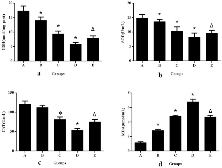
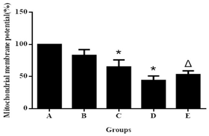
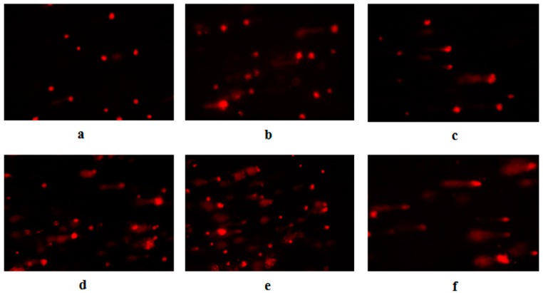
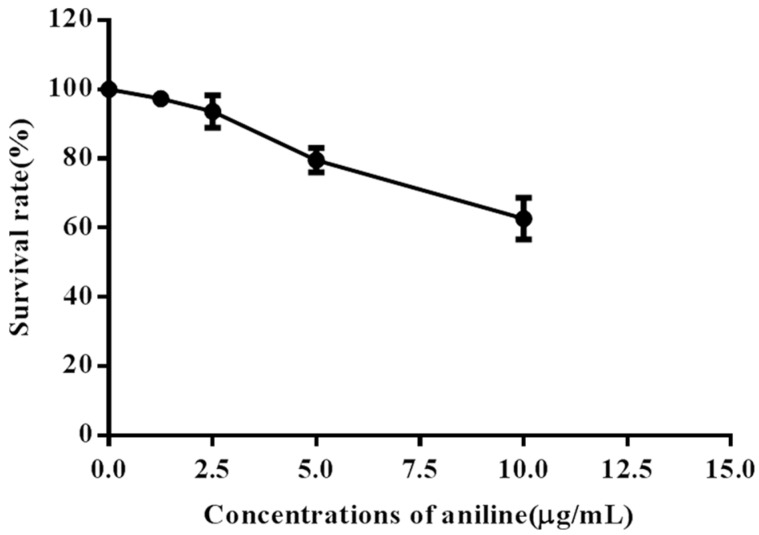
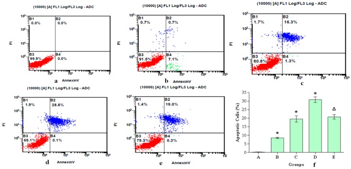
The toxicity and carcinogenicity of aniline in humans and animals have been well documented. However, the molecular mechanism involved in aniline-induced liver toxicity and carcinogenesis remains unclear. In our research, primary cultured hepatocytes were exposed to aniline (0, 1.25, 2.50, 5.0 and 10.0 μg/mL) for 24 h in the presence or absence of N-acetyl-l-cysteine (NAC). Levels of reactive oxygen species (ROS), malondialdehyde (MDA), and glutathione (GSH), activities of superoxide dismutase (SOD) and catalase (CAT), mitochondrial membrane potential, DNA damage, cell viability, and apoptosis were detected. Levels of ROS and MDA were significantly increased and levels of GSH and CAT, activity of SOD, and mitochondrial membrane potential in hepatocytes were significantly decreased by aniline compared with the negative control group. The tail moment and DNA content of the tail in exposed groups were significantly higher than those in the negative control group. Cell viability was reduced and apoptotic death was induced by aniline in a concentration-dependent manner. The phenomena of ROS generation, oxidative damage, loss of mitochondrial membrane potential, DNA damage and apoptosis could be prevented if ROS inhibitor NAC was added. ROS generation is involved in the loss of mitochondrial membrane potential and DNA injury, which may play a role in aniline-induced apoptosis in hepatocytes. Our study provides insight into the mechanism of aniline-induced toxicity and apoptosis of hepatocytes.
Keywords: aniline; apoptosis; hepatocytes; reactive oxygen species.
Conflict of interest statement
The authors declare no conflict of interest.
Figures
Similar articles
-
Roles of reactive oxygen species and mitochondria in cadmium-induced injury of liver cells.Toxicol Ind Health. 2011 Apr;27(3):249-56. doi: 10.1177/0748233710386408. Epub 2010 Dec 9. Toxicol Ind Health. 2011. PMID: 21148202
-
[Study on the molecular mechanism of autophagy and apoptosis induced by ultrafine carbon black in human bronchial epithelial cells and the intervention effect of N-acetylcysteine].Zhonghua Lao Dong Wei Sheng Zhi Ye Bing Za Zhi. 2024 Sep 20;42(9):656-667. doi: 10.3760/cma.j.cn121094-20231010-00080. Zhonghua Lao Dong Wei Sheng Zhi Ye Bing Za Zhi. 2024. PMID: 39394703 Chinese.
-
Oxidative stress is involved in Dasatinib-induced apoptosis in rat primary hepatocytes.Toxicol Appl Pharmacol. 2012 Jun 15;261(3):280-91. doi: 10.1016/j.taap.2012.04.010. Epub 2012 Apr 17. Toxicol Appl Pharmacol. 2012. PMID: 22538170
-
[A pathophysiological role of cytochrome p450 involved in production of reactive oxygen species].Yakugaku Zasshi. 2013;133(4):435-50. doi: 10.1248/yakushi.12-00263. Yakugaku Zasshi. 2013. PMID: 23546588 Review. Japanese.
-
Oxidative stress promotes the regression of fetal liver hemopoiesis.Biochemistry (Mosc). 2004 Jan;69(1):18-22, 1 p following 74. doi: 10.1023/b:biry.0000016346.61403.24. Biochemistry (Mosc). 2004. PMID: 14972013 Review.
Cited by
-
The Novel Antipsychotic Lumateperone (Iti-007) in the Treatment of Schizophrenia: A Systematic Review.Brain Sci. 2023 Nov 26;13(12):1641. doi: 10.3390/brainsci13121641. Brain Sci. 2023. PMID: 38137089 Free PMC article. Review.
-
Application of in vitro Drug Metabolism Studies in Chemical Structure Optimization for the Treatment of Fibrodysplasia Ossificans Progressiva (FOP).Front Pharmacol. 2019 Apr 24;10:234. doi: 10.3389/fphar.2019.00234. eCollection 2019. Front Pharmacol. 2019. PMID: 31068801 Free PMC article.
-
Exploring the Critical Factors Limiting Polyaniline Biocompatibility.Polymers (Basel). 2019 Feb 19;11(2):362. doi: 10.3390/polym11020362. Polymers (Basel). 2019. PMID: 30960346 Free PMC article.
-
Bioisosteric Modification of To042: Synthesis and Evaluation of Promising Use-Dependent Inhibitors of Voltage-Gated Sodium Channels.ChemMedChem. 2021 Dec 6;16(23):3588-3599. doi: 10.1002/cmdc.202100496. Epub 2021 Oct 5. ChemMedChem. 2021. PMID: 34519427 Free PMC article.
-
Hybrid non-animal modeling: A mechanistic approach to predict chemical hepatotoxicity.J Hazard Mater. 2024 Jun 5;471:134297. doi: 10.1016/j.jhazmat.2024.134297. Epub 2024 Apr 12. J Hazard Mater. 2024. PMID: 38677119 Free PMC article.
References
MeSH terms
Substances
LinkOut - more resources
Full Text Sources
Other Literature Sources
Miscellaneous
