

Visualization of Neutrophil Extracellular Traps and Fibrin Meshwork in Human Fibrinopurulent Inflammatory Lesions: III. Correlative Light and Electron Microscopic Study
- PMID: 27917008
- PMCID: PMC5130345
- DOI: 10.1267/ahc.16028
Visualization of Neutrophil Extracellular Traps and Fibrin Meshwork in Human Fibrinopurulent Inflammatory Lesions: III. Correlative Light and Electron Microscopic Study
Abstract
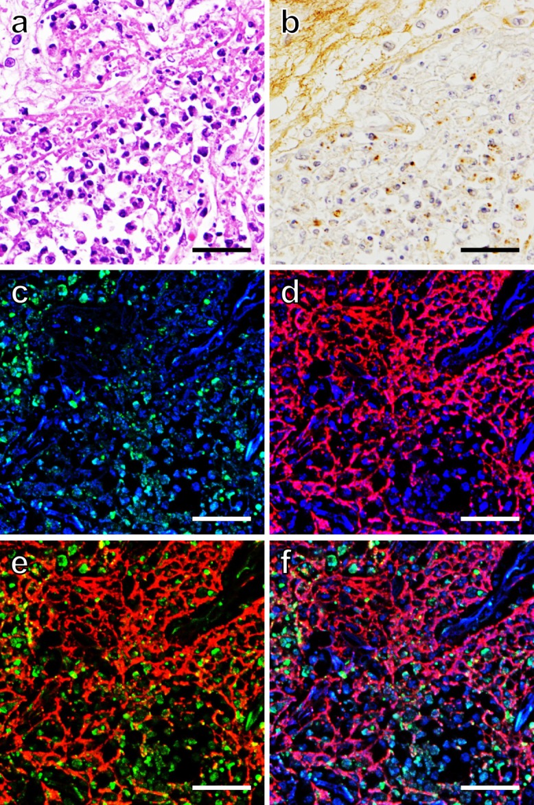
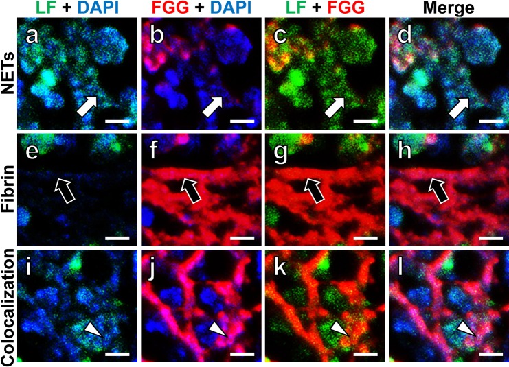
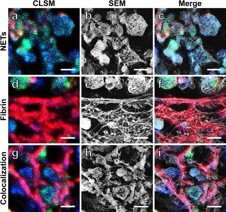
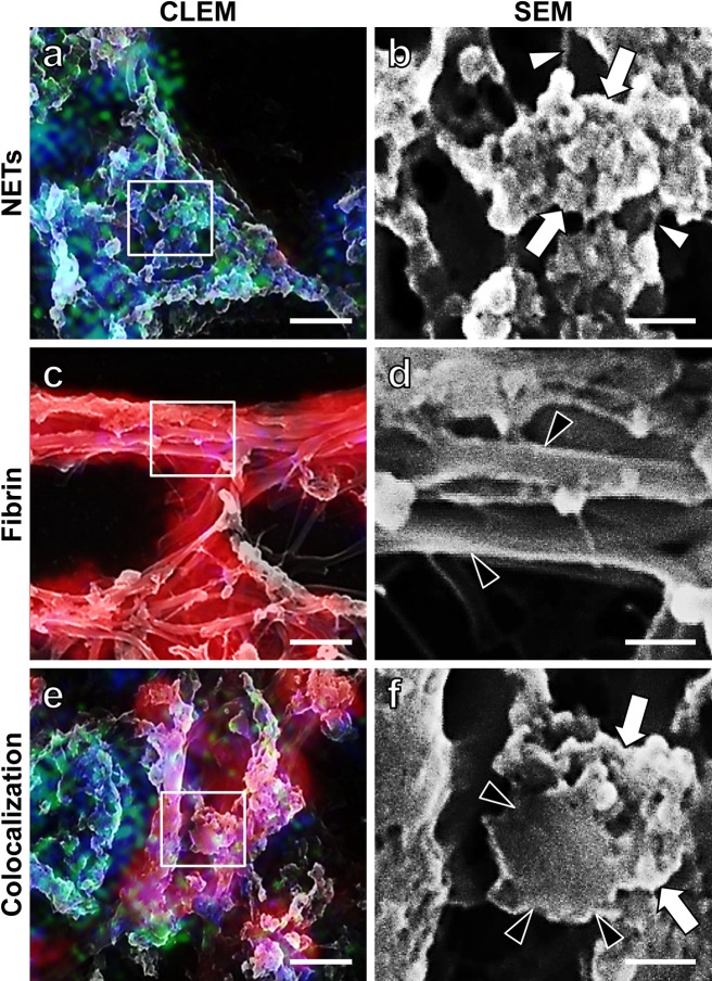
Neutrophil extracellular traps (NETs) released from dead neutrophils at the site of inflammation represent webs of neutrophilic DNA stretches dotted with granule-derived antimicrobial proteins, including lactoferrin, and play important roles in innate immunity against microbial infection. We have shown the coexistence of NETs and fibrin meshwork in varied fibrinopurulent inflammatory lesions at both light and electron microscopic levels. In the present study, correlative light and electron microscopy (CLEM) employing confocal laser scanning microscopy and scanning electron microscopy was performed to bridge light and electron microscopic images of NETs and fibrin fibrils in formalin-fixed, paraffin-embedded, autopsied lung sections of legionnaire's pneumonia. Lactoferrin immunoreactivity and 4'-6-diamidino-2-phenylindole (DAPI) reactivity were used as markers of NETs, and fibrin was probed by fibrinogen gamma chain. Of note is that NETs light microscopically represented as lactoferrin and DAPI-colocalized dots, 2.5 μm in diameter. CLEM gave super-resolution images of NETs and fibrin fibrils: "Dotted" NETs were ultrastructurally composed of fine filaments and masses of 58 nm-sized globular materials. A fibrin fibril consisted of clusters of smooth-surfaced filaments. NETs filaments (26 nm in diameter) were significantly thinner than fibrin filaments (295 nm in diameter). Of note is that CLEM was applicable to formalin-fixed, paraffin-embedded sections of autopsy material.
Keywords: confocal laser scanning microscopy; correlative light and electron microscopy; fibrin; neutrophil extracellular traps; scanning electron microscopy.
Figures
Similar articles
-
Visualization of Neutrophil Extracellular Traps and Fibrin Meshwork in Human Fibrinopurulent Inflammatory Lesions: II. Ultrastructural Study.Acta Histochem Cytochem. 2016 Aug 30;49(4):117-23. doi: 10.1267/ahc.16016. Epub 2016 Jul 30. Acta Histochem Cytochem. 2016. PMID: 27682015 Free PMC article.
-
Visualization of Neutrophil Extracellular Traps and Fibrin Meshwork in Human Fibrinopurulent Inflammatory Lesions: I. Light Microscopic Study.Acta Histochem Cytochem. 2016 Aug 30;49(4):109-16. doi: 10.1267/ahc.16015. Epub 2016 Jul 30. Acta Histochem Cytochem. 2016. PMID: 27682014 Free PMC article.
-
Fibrin mimics neutrophil extracellular traps in SEM.Ultrastruct Pathol. 2010 Aug;34(4):226-31. doi: 10.3109/01913121003725721. Ultrastruct Pathol. 2010. PMID: 20594043
-
How Neutrophil Extracellular Traps Become Visible.J Immunol Res. 2016;2016:4604713. doi: 10.1155/2016/4604713. Epub 2016 May 16. J Immunol Res. 2016. PMID: 27294157 Free PMC article. Review.
-
The pathway of neutrophil extracellular traps towards atherosclerosis and thrombosis.Atherosclerosis. 2019 Sep;288:9-16. doi: 10.1016/j.atherosclerosis.2019.06.919. Epub 2019 Jun 29. Atherosclerosis. 2019. PMID: 31280097 Review.
Cited by
-
Visualization and Quantification of Neutrophil Extracellular Traps.Methods Mol Biol. 2021;2255:87-95. doi: 10.1007/978-1-0716-1162-3_9. Methods Mol Biol. 2021. PMID: 34033097 Free PMC article.
-
Morphologic Analysis of M2 Macrophage in Glioblastoma: Involvement of Macrophage Extracellular Traps (METs).Acta Histochem Cytochem. 2022 Aug 27;55(4):111-118. doi: 10.1267/ahc.22-00018. Epub 2022 Aug 10. Acta Histochem Cytochem. 2022. PMID: 36060293 Free PMC article.
-
Challenges and insights in detecting early inflammation in DSS-induced colitis using chemiluminescence.Photochem Photobiol Sci. 2025 May;24(5):765-777. doi: 10.1007/s43630-025-00724-9. Epub 2025 May 1. Photochem Photobiol Sci. 2025. PMID: 40312581
-
Serum amyloid A binds to fibrin(ogen), promoting fibrin amyloid formation.Sci Rep. 2019 Feb 28;9(1):3102. doi: 10.1038/s41598-019-39056-x. Sci Rep. 2019. PMID: 30816210 Free PMC article.
-
Correlative Light-Electron Microscopy detects lipopolysaccharide and its association with fibrin fibres in Parkinson's Disease, Alzheimer's Disease and Type 2 Diabetes Mellitus.Sci Rep. 2018 Nov 14;8(1):16798. doi: 10.1038/s41598-018-35009-y. Sci Rep. 2018. PMID: 30429533 Free PMC article.
References
-
- Beiter K., Wartha F., Albiger B., Normark S., Zychlinsky A. and Henriques-Normark B. (2006) An endonuclease allows Streptococcus pneumoniae to escape from neutrophil extracellular traps. Curr. Biol. 16; 401–407. - PubMed
-
- Brinkmann V., Reichard U., Goosmann C., Fauler B., Uhlemann Y., Weiss D. S., Weinrauch Y. and Zychlinsky A. (2004) Neutrophil extracellular traps kill bacteria. Science 303; 1532–1535. - PubMed
-
- Buchanan J. T., Simpson A. J., Aziz R. K., Liu G. Y., Kristian S. A., Kotb M., Feramisco J. and Nizet V. (2006) DNase expression allows the pathogen group A Streptococcus to escape killing in neutrophil extracellular traps. Curr. Biol. 16; 396–400. - PubMed
-
- Chung C. L., Chen Y. C. and Chang S. C. (2003) Effect of repeated thoracenteses on fluid characteristics, cytokines, and fibrinolytic activity in malignant pleural effusion. Chest 123; 1188–1195. - PubMed
LinkOut - more resources
Full Text Sources
Other Literature Sources