. 2016 Dec 1;72(Pt 12):1298-1307.
doi: 10.1107/S2059798316018623. Epub 2016 Nov 30.

Structural analysis of the bright monomeric yellow-green fluorescent protein mNeonGreen obtained by directed evolution

Affiliations

Structural analysis of the bright monomeric yellow-green fluorescent protein mNeonGreen obtained by directed evolution

Damien Clavel et al. Acta Crystallogr D Struct Biol. .

Abstract

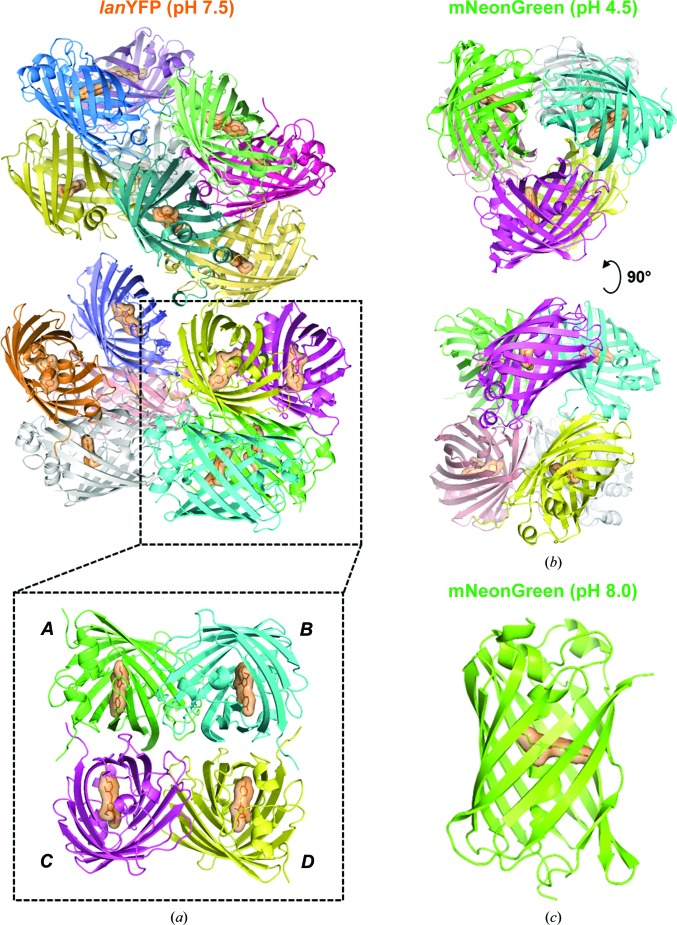
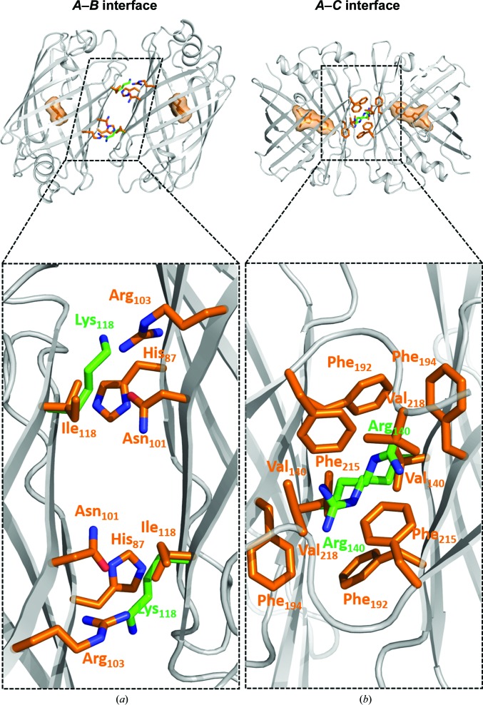
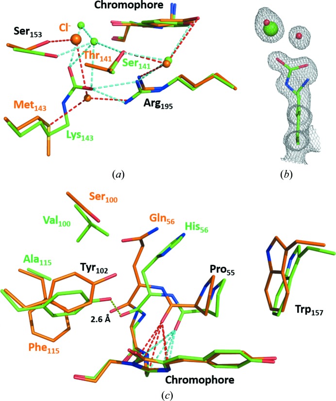
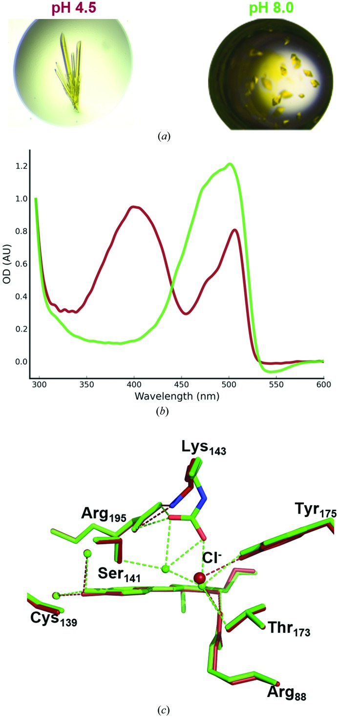
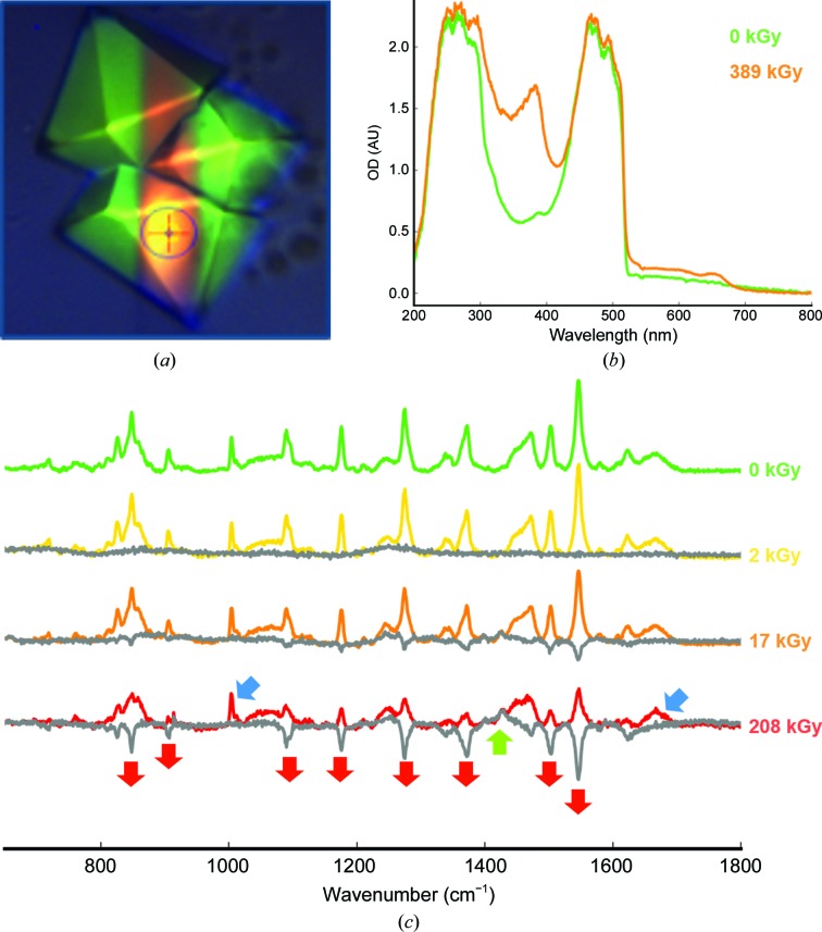
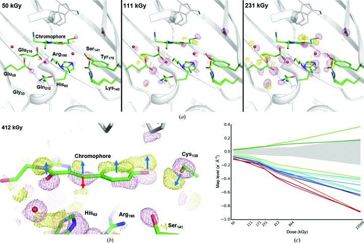
Until recently, genes coding for homologues of the autofluorescent protein GFP had only been identified in marine organisms from the phyla Cnidaria and Arthropoda. New fluorescent-protein genes have now been found in the phylum Chordata, coding for particularly bright oligomeric fluorescent proteins such as the tetrameric yellow fluorescent protein lanYFP from Branchiostoma lanceolatum. A successful monomerization attempt led to the development of the bright yellow-green fluorescent protein mNeonGreen. The structures of lanYFP and mNeonGreen have been determined and compared in order to rationalize the directed evolution process leading from a bright, tetrameric to a still bright, monomeric fluorescent protein. An unusual discolouration of crystals of mNeonGreen was observed after X-ray data collection, which was investigated using a combination of X-ray crystallography and UV-visible absorption and Raman spectroscopies, revealing the effects of specific radiation damage in the chromophore cavity. It is shown that X-rays rapidly lead to the protonation of the phenolate O atom of the chromophore and to the loss of its planarity at the methylene bridge.

Keywords: Branchiostoma lanceolatum; fluorescent protein; in crystallo optical spectroscopy; online Raman spectroscopy; specific radiation damage.


Figures

Figure 1
Figure 1
Asymmetric unit in the various crystal forms of lanYFP and mNeonGreen. (a) lanYFP tetramer of tetramers in crystals of space group P21 obtained at pH 7.5. (b) mNeonGreen dimer of trimers in crystals of space group P212121 obtained at pH 4.5. (c) mNeonGreen monomer in crystals of space group P6522 obtained at pH 8.0.
Figure 2
Figure 2
(a) AB and (b) AC interfaces in lanYFP (orange side chains) featuring two key mutations ensuring disruption of the interactions in mNeonGreen (green side chains).
Figure 3
Figure 3
Comparison of the environment of the chromophore in lanYFP (orange) and in mNeonGreen (green) at pH 8.0. (a) Close-up of the chloride-binding site in lanYFP and the carboxylated lysine in mNeonGreen at physiological pH. (b) σA-weighted 2F oF c electron-density map contoured at a 1.0σ level around Lys143 in mNeonGreen, the major conformation of which is carboxylated and the minor conformation of which allows the binding of a chloride ion. (c) Close-up of the differences located on the other side of the chromophore. The strong hydrogen bond between Tyr102 and the carbonyl group of His56 in mNeonGreen is represented as a yellow dashed line. The lone pair–π interaction between the carbonyl group of Pro55 and the imidazolinone ring of the chromophore is represented as red dashed lines in lanYFP and cyan dashed lines in mNeonGreen
Figure 4
Figure 4
Spectroscopic and structural comparison of mNeonGreen at acidic and near-physiological pH values. (a) mNeonGreen crystal morphologies obtained at pH 4.5 (top) and pH 8.0 (bottom). (b) UV–visible absorption spectra of mNeonGreen crystals at pH 4.5 (dark red) and pH 8.0 (green). (c) Superposition of the chromophore environment in mNeonGreen at pH 4.5 (dark red) and pH 8.0 (green).
Figure 5
Figure 5
X-ray-induced spectroscopic changes of mNeonGreen. (a) Picture of mNeonGreen crystals after a 730 kGy X-ray data collection. (b) UV–visible absorption spectra of the irradiated (orange trace) and non-irradiated (green trace) areas of an mNeonGreen crystal. (c) Series of online Raman spectra measured on an mNeonGreen crystal with increasing X-ray dose. Grey traces correspond to spectra subtracted for the zero-dose spectrum. Blue arrows indicate invariant peaks characteristics of proteins, red arrows indicate decreasing peaks and the green arrow indicates an increasing peak.
Figure 6
Figure 6
X-ray-induced structural changes in the chromophore cavity of mNeonGreen. (a) Fourier difference maps F o(n) − F o(1) at increasing X-ray doses contoured at a 6.0σ level (pink, negative; yellow, positive). (b) Close-up on the chromophore at a 4.0σ level after 412 kGy. Dark blue arrows indicate the upward movement of the whole chromophore and the opposite movement of Cys139. The red arrow indicates the concomitant formation of a kinked configuration of the chromophore upon loss of conjugation at the exocyclic linkage. (c) X-ray-induced electron loss or gain displayed as a function of X-­ray dose on a logarithmic scale. Three ensembles of affected groups can be distinguished: damage to Glu35, Glu210 and the water in between is represented in red, damage to the methylene bridge in green and strong damage to other groups listed in Table 3 ▸ in blue. The noise level (corresponding to peak heights with a σ level between −3.0 and +3.0) at each dose is represented by a light grey shade.

References

    1. Adam, V., Carpentier, P., Violot, S., Lelimousin, M., Darnault, C., Nienhaus, G. U. & Bourgeois, D. (2009). J. Am. Chem. Soc. 131, 18063–18065. - PubMed
    1. Alieva, N. O., Konzen, K. A., Field, S. F., Meleshkevitch, E. A., Hunt, M. E., Beltran-Ramirez, V., Miller, D. J., Wiedenmann, J., Salih, A. & Matz, M. V. (2008). PLoS One, 3, e2680. - PMC - PubMed
    1. Bell, A. F., He, X., Wachter, R. M. & Tonge, P. J. (2000). Biochemistry, 39, 4423–4431. - PubMed
    1. Bui, S., von Stetten, D., Jambrina, P. G., Prangé, T., Colloc’h, N., de Sanctis, D., Royant, A., Rosta, E. & Steiner, R A. (2014). Angew. Chem. Int. Ed. Engl. 53, 13710–13714. - PMC - PubMed
    1. Burmeister, W. P. (2000). Acta Cryst. D56, 328–341. - PubMed

Publication types

Substances