. 2016 Dec 5:6:38253.
doi: 10.1038/srep38253.

Incomplete and delayed Sox2 deletion defines residual ear neurosensory development and maintenance

Affiliations

Incomplete and delayed Sox2 deletion defines residual ear neurosensory development and maintenance

Martina Dvorakova et al. Sci Rep. .

Abstract

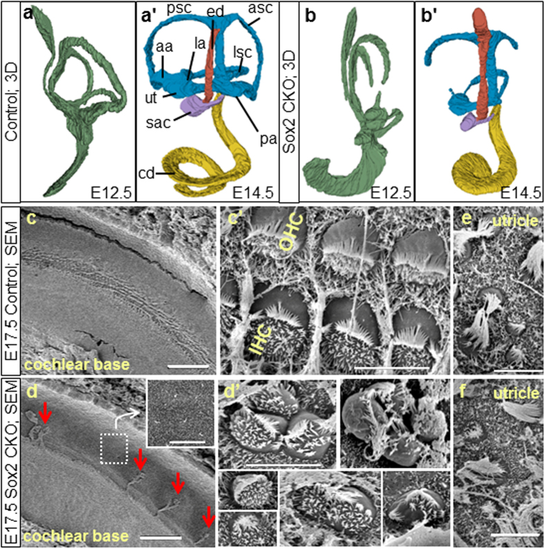
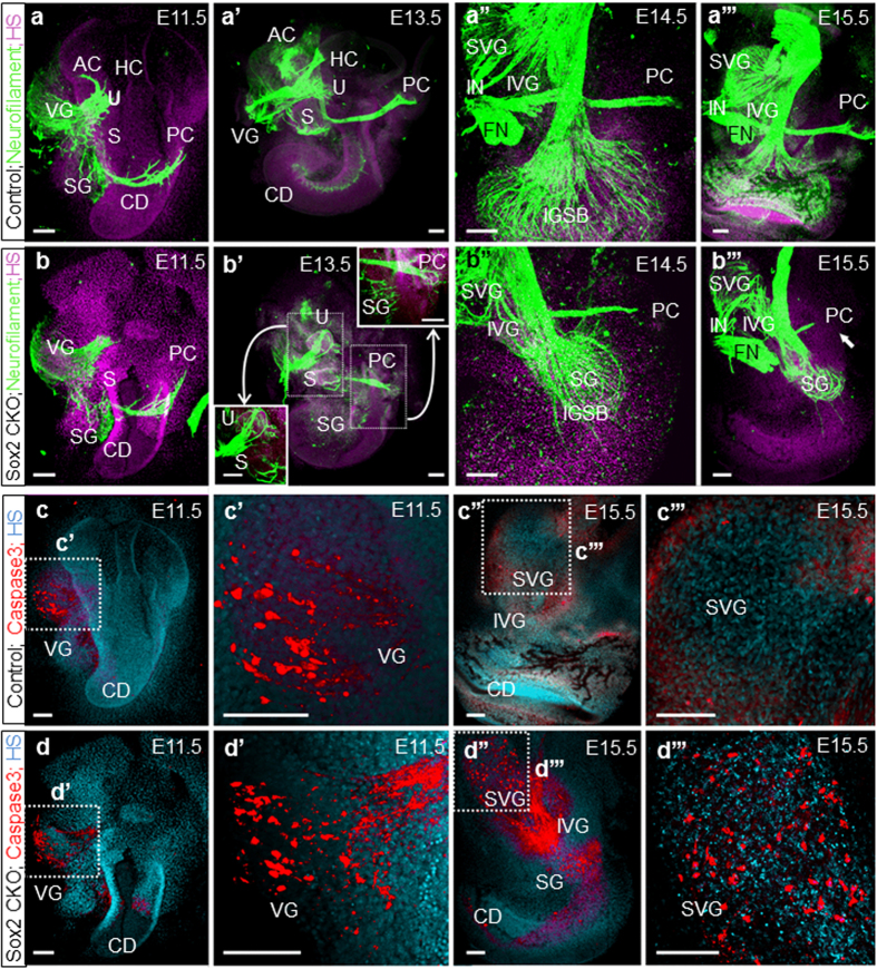
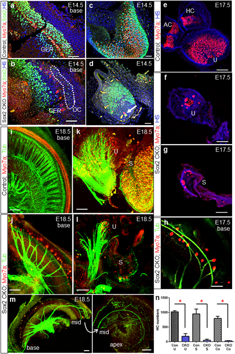
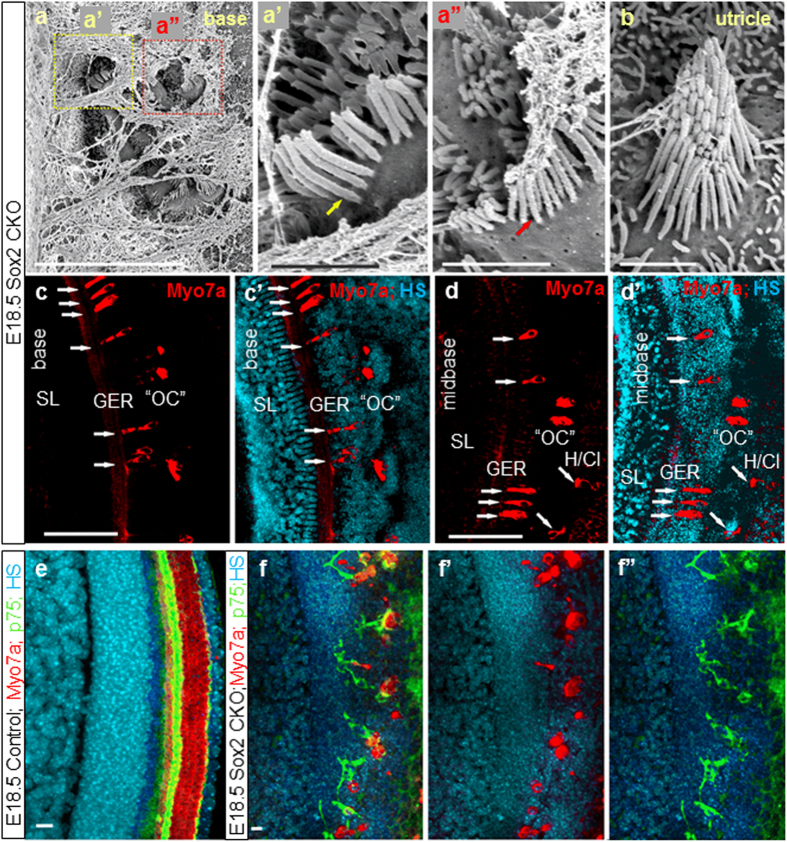
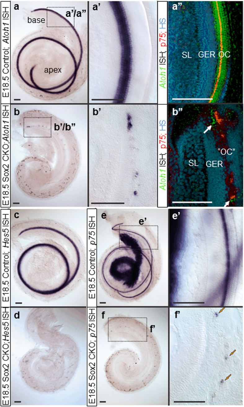
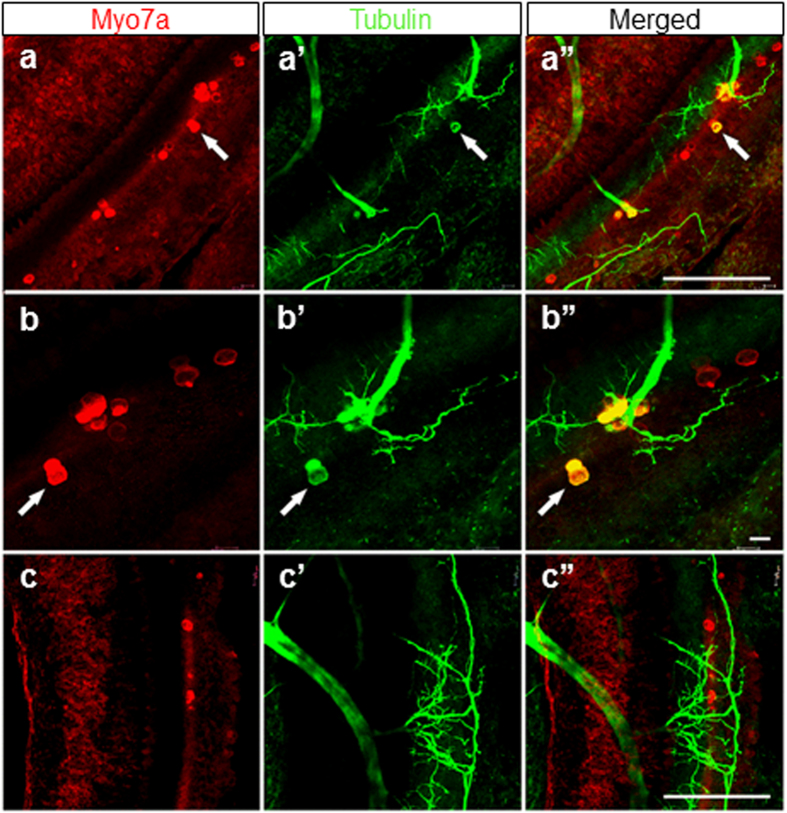
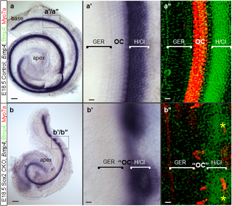
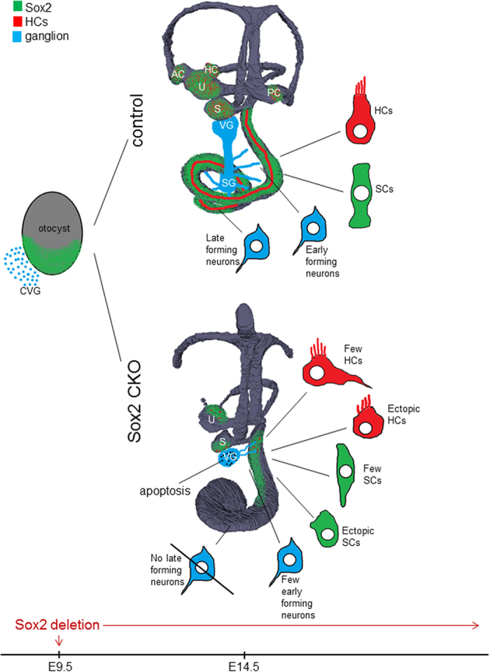
The role of Sox2 in neurosensory development is not yet fully understood. Using mice with conditional Islet1-cre mediated deletion of Sox2, we explored the function of Sox2 in neurosensory development in a model with limited cell type diversification, the inner ear. In Sox2 conditional mutants, neurons initially appear to form normally, whereas late- differentiating neurons of the cochlear apex never form. Variable numbers of hair cells differentiate in the utricle, saccule, and cochlear base but sensory epithelium formation is completely absent in the apex and all three cristae of the semicircular canal ampullae. Hair cells differentiate only in sensory epithelia known or proposed to have a lineage relationship of neurons and hair cells. All initially formed neurons lacking hair cell targets die by apoptosis days after they project toward non-existing epithelia. Therefore, late neuronal development depends directly on Sox2 for differentiation and on the survival of hair cells, possibly derived from common neurosensory precursors.


Conflict of interest statement

The authors declare no competing financial interests.

Figures

Figure 1
Figure 1. Altered morphology of the Sox2 CKO inner ear.
(a-b’) 3D-reconstruction reveals severe changes in the developing inner ear at E12.5 and E14.5. (b,b’) No ampullae of semicircular canals and only rudiments of the posterior and anterior semicircular canals are present in Sox2 CKO. The utricle and saccule are smaller. (a’,b’) The cochlear duct (cd) has decreased coiling and is shorter compared to controls. (cf) Scanning electron microscopy shows a few individual cells and small clumps of cells with a hair cell-like phenotype in the base of the Sox2 CKO cochlea (arrows). (d) The rest of the organ of Corti is missing as shown by the overview of the whole cd width and by magnification of the sensory epithelium area. (d’) HCs vary in size, orientation and bundle organization. (e,f) The cellular phenotype of differentiated HCs in the Sox2 CKO utricle is comparable to controls. aa, anterior ampulla; asc, anterior semicircular canal; cd, cochlear duct; ed, endolymphatic duct; la, lateral ampulla; lsc, lateral semicircular canal; pa, posterior ampulla; psc, posterior semicircular canal; sac, saccule; ut, utricle; OHC, outer hair cells; IHC, inner hair cells. Scale bars: 50 μm (c,d), 5 μm (c’,d’,e,f).
Figure 2
Figure 2. Isl1-cre mediated Sox2 loss disrupts neuron formation and results in massive neuronal degeneration by activation of Caspase3.
(a,b) Immunofluorescence staining of neurofilament in the Sox2 CKO shows similar formation of vestibular neurons at E11.5 compared to controls. (a’,b’) At E13.5, fibers are aberrantly projecting toward the utricle (or combined utricle and anterior and horizontal canal cristae) and posterior canal cristae of the Sox2 CKO. (a”,b”,a”’,b”’) Fibers to the posterior canal crista start to retract in the absence of target HCs starting at E14.5 in the mutant. (b”’) Only a few radial fibers are formed near the base of the E15.5 Sox2 CKO cochlea. (c, c’,d,d’) Immunofluorescence of activated Caspase3 reveals positive staining restricted mainly in the VG in the E11.5 Sox2 CKO comparable to the control littermates. (c”,c”’,d”,d”’) However, Caspase3 mediated cell death is massively progressed to IVG, SVG, and SG at E15.5 compared to no caspase positive cells in the control littermates. Scale bars: 100 μm. AC, anterior canal crista; CD, cochlear duct; FN, facial nerve; HS, Hoechst nuclear stain; IGSB, intraganglionic spiral bundle; IVG, inferior vestibular ganglion; IN, intermediate nerve; HC, horizontal canal crista; PC, posterior canal crista; S, saccule; SG, spiral ganglion; SVG, superior vestibular ganglion; U, utricle; VG, vestibular ganglia.
Figure 3
Figure 3. Delayed deletion of Sox2 results in the differentiation of some neurosensory cells in the basal cochlear turn and in the vestibular organs.
(a,b) Sox2+ cells in the Sox2 CKO cochlea are detected only in the base at the age of E14.5 and disappear later in development. (b) The strong Sox2 expression domain is shifted toward the GER. Similarly, Myo7a+ cells do not differentiate in the proper area of OC. Some weak Sox2 expression remains in the OC area of Sox2 CKO (dotted area). (c,d) Variable numbers of HCs (Myo7a+) and supporting cells (Sox2+) develop in the Sox2 CKO vestibular system. (d) HCs in the saccule also develop in the area that lacks supporting cells (arrow). (e–h) Some poorly differentiated Myo7a+ HCs are present in the utricle, saccule and basal turn of the cochlea of the Sox2 CKO at E17.5. (i–m) At E18.5, the innervation of mutant cochlea, saccule and utricle is severely reduced and shows an unusual pattern compared to controls. Fibers show mostly directional growth toward remaining HCs but also transient expansion into HC-free regions. (n) The quantification of Myo7a positive HCs after whole mount immunostaining shows a striking reduction of HCs in the Sox2 CKO inner ear compared to littermate controls for the utricle (U), saccule (S) and cochlea (Co). Myo7a+ HCs were counted after whole mount immunostaining using LAS AF Lite draw counter to avoid counting error. The total number of HCs was determined in the entire utricle and saccule, and in the entire Sox2 CKO cochlea. The number of HCs in the control cochlea represents the total number of HCs in 1.5 mm of the base. The values represent means ± SD (N = 4–7 individuals/group). *P < 0.0001, t-test. Scale bars: 50 μm (a–h), 100 μm (i–m). AC, anterior crista; GER, greater epithelial ridge; HC, horizontal crista; OC, organ of Corti; S, saccule; U, utricle.
Figure 4
Figure 4. Aberrant HCs in ectopic topology and abnormal pillar cells are formed in the Sox2 CKO.
Patches of HCs in the base of the E18.5 Sox2 CKO cochlea are covered by a tectorial membrane with HCs in the topology of inner HCs displaying both large diameter (a,a’) and small diameter (a,a”) stereocilia reminiscent of inner and outer HCs, respectively. (b) Vestibular HCs show normal organization of stereocilia but many display variability in stereocilia diameter in a single HC, normally associated with either type I or type II vestibular HCs. (c-d’) Scattered Myo7a positive HCs are detected in the area corresponding topologically to the organ of Corti (OC); however, forming atypical organ of Corti (“OC”) in the mutant. Immunostaining of Myo7a reveals formation of HCs in the ectopic topologies, medial to OC, in the GER, as well as lateral to OC (in the area of Hensen/Claudius cells) (white arrows) in addition to the area of “OC” in the E18.5 Sox2 CKO. (e-f”) The combination of p75 and Myo7a immunolabeling shows an unusual configuration and distribution of p75 positive cells near the remaining Myo7a positive HCs in E18.5 Sox2 CKO compared to the single row of p75+ inner pillar cells in control littermates (e). Scale bars: 10 μm (a,e-f”), 1 μm (a’,a”,b), 100 μm (c-d’). GER, greater epithelial ridge; H/Cl, Hensen/Claudius cells; OC, organ of Corti; “OC”, atypical organ of Corti in the mutant; SL, spiral limbus.
Figure 5
Figure 5. Loss of Sox2 affects downstream gene expression in the organ of Corti.
(a,a’,b,b’) Atoh1 expression is dramatically reduced in Sox2 CKO mice at E18.5. (a”,b”) Atoh1 ISH signal converting into a fluorescent signal shows the relative topology to the inner pillar cell marker p75. Note that the scattered HCs are found medial and lateral to p75 positive cells (white arrows; b”). However, p75 expression (arrows) is discontinuous in the base (b”,f,f’) and absent in the apex compared to prominent labeling in the inner pillar cells and the spiral ganglion neurons in control animals (e,e’). (c,d) Another supporting cell marker, Hes5, shows no expression at all in the Sox2 CKO mice at E18.5. Scale bars: 100 μm. GER, greater epithelial ridge; OC, organ of Corti; “OC”, atypical organ of Corti in the mutant; SL, spiral limbus.
Figure 6
Figure 6. Some HCs are positive for both Myo7a and tubulin.
These single or groups of Myo7a positive HCs of E18.5 Sox2 CKO mice show a patchy distribution (a,b,c) and an unusual pattern of innervation (a’,b’,c’). Note that most fibers are targeted toward Myo7a positive HCs, others are sometimes widely distributed in the topological equivalent of the organ of Corti. (a”,b”,c”) Some Myo7a positive cells are also positive for antibody directed against tubulin, normally a reliable neuronal marker in the ear. Scale bars: 100 μm, except b,b” that indicates 10 μm.
Figure 7
Figure 7. The cellular boundaries of the inner ear are changed in the Sox2 CKO.
(a,a’,b,b’) Loss of Sox2 results in aberration of Bmp4 expression. Instead of being separated by the organ of Corti from the GER, Bmp4 expression is adjacent to the GER. (b, b’,b”) Only the base shows rings of lateral Bmp4 expression and Myo7a positive HCs are both in the center of these rings as well as at the boundary between GER and Bmp4 domain (b”; yellow asterisks). (a’-a”) In controls, the Bmp4 expression in Hensen/Claudius cells is always lateral to the organ of Corti. Scale bars: 10 μm except 100 μm in a and b. GER, greater epithelial ridge; H/Cl; Hensen/Claudius cells; OC, organ of Corti; “OC”, atypical organ of Corti in the mutant.
Figure 8
Figure 8. Summary of Sox2 CKO inner ear changes.
Sox2 deletion by Isl1-cre results in profound morphological changes at E14.5: all three cristae of the semicircular canal ampullae are missing, all remaining sensory organs are smaller and have a decreased size of the sensory area. Neuronal formation is eliminated in the apex of the cochlea, whereas vestibular and basal turn neurons gradually die due to limited support by the reduced sensory epithelia. Loss of innervation toward all epithelia except for the apex of the cochlea is secondary to the lack or reduced differentiation of HCs, which is either completely absent in the apex and semicircular canal cristae, or variably disabled in the base of the cochlea, utricle, and saccule. Many HCs have an unusual neurosensory phenotype. Similarly, supporting cells (SCs) have atypical features, altered expression of markers, and abnormal distribution, forming the aberrant organ of Corti. Many ectopic HCs and SCs are also found in the cochlear base. The spatial distribution of Sox2 expression is shown in green, blue color shows neurons, and red depicts HCs. AC, anterior crista; CVG, cochleovestibular ganglion; HC, horizontal crista; HCs, hair cells; PC, posterior crista; S, saccule; SCs, supporting cells; SG, spiral ganglion; U, utricle; VG, vestibular ganglion.

References

    1. Kondoh H. & Lovell-Badge R. Sox2: Biology and Role in Development and Disease. 3–15 (Elsevier, 2016).
    1. Reiprich S. & Wegner M. From CNS stem cells to neurons and glia: Sox for everyone. Cell Tissue Res 359, 111–124 (2015). - PubMed
    1. Telley L. et al.. Sequential transcriptional waves direct the differentiation of newborn neurons in the mouse neocortex. Science 351, 1443–1446 (2016). - PubMed
    1. Fritzsch B., Jahan I., Pan N. & Elliott K. L. Evolving gene regulatory networks into cellular networks guiding adaptive behavior: an outline how single cells could have evolved into a centralized neurosensory system. Cell Tissue Res 359, 295–313 (2015). - PMC - PubMed
    1. Fritzsch B., Pan N., Jahan I. & Elliott K. L. Inner ear development: building a spiral ganglion and an organ of Corti out of unspecified ectoderm. Cell Tissue Res 368, 7–24 (2015). - PMC - PubMed

Publication types

Substances

LinkOut - more resources