. 2017 Jan 9;56(2):524-529.
doi: 10.1002/anie.201609427. Epub 2016 Dec 5.

Development of Cell-Permeable, Non-Helical Constrained Peptides to Target a Key Protein-Protein Interaction in Ovarian Cancer

Affiliations

Development of Cell-Permeable, Non-Helical Constrained Peptides to Target a Key Protein-Protein Interaction in Ovarian Cancer

Mareike M Wiedmann et al. Angew Chem Int Ed Engl. .

Abstract

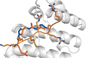
There is a lack of current treatment options for ovarian clear cell carcinoma (CCC) and the cancer is often resistant to platinum-based chemotherapy. Hence there is an urgent need for novel therapeutics. The transcription factor hepatocyte nuclear factor 1β (HNF1β) is ubiquitously overexpressed in CCC and is seen as an attractive therapeutic target. This was validated through shRNA-mediated knockdown of the target protein, HNF1β, in five high- and low-HNF1β-expressing CCC lines. To inhibit the protein function, cell-permeable, non-helical constrained proteomimetics to target the HNF1β-importin α protein-protein interaction were designed, guided by X-ray crystallographic data and molecular dynamics simulations. In this way, we developed the first reported series of constrained peptide nuclear import inhibitors. Importantly, this general approach may be extended to other transcription factors.

Keywords: constrained peptides; drug discovery; nuclear import; peptide therapeutics; peptidomimetics.


Figures

Figure 1
Figure 1
Proposed scheme for targeting the nuclear import of HNF1β through the HNF1β–importin α PPI: 1) The IBB domain of importin α binds to importin β to free up the NLS‐binding sites on importin α. 2) HNF1β NLS recognition by a heterodimeric complex composed of importin α and importin β, 3, 4) To enable HNF1β to be imported in the nucleus, the HNF1β NLS has to bind to the importin α–β heterodimer. The constrained peptide competes for this binding, thereby impairing the import of HNF1β. 5) Release of the constrained peptide through RanGTP binding to importin β. Reproduced and modified from Kobe et al.16
Figure 2
Figure 2
Relative proliferation of PEO1, JHOC5, JHOC7, JHOC9, OVISE, and SKOV3 CCC lines with n=4 after HNF1β shRNA knockdown. The mean is shown, with error bars showing the SEM. Statistical significance was assessed with multiple t‐tests and the Holm–Šídák method with α=5 %. Optical densities (ODs) are given relative to their respective non‐target knockdown OD value and background OD was subtracted. Only shRNA knockdown clone 583 at 96 h was considered here. * indicates P<0.02.
Figure 3
Figure 3
Binding interactions of HNF1β NLS peptide (orange) with mImportin α1 (gray) determined from X‐ray crystallography and MD simulations. The trajectory structures shown are final snapshots taken from the end of the simulations. A) The backbone carbonyl oxygen of Thr1 hydrogen bonds to the side chain of Arg238 in the obtained crystal structure (PDB ID: 5K9S). B) The side chain of Thr1 hydrogen bonds with the side chain of Asp270 in the MD simulations. C) Arg9 forms a salt bridge with Glu465 from a neighboring protein chain (pink) in the crystal structure. D) Arg9 forms a salt bridge with Glu107 in the MD simulations.
Figure 4
Figure 4
Energetic analysis of the MD simulations of the complex of HNF1β NLS peptide with mImportin α1. A) Binding free energy contributions of HNF1β NLS peptide residues. B) Computational alanine scanning of HNF1β NLS peptide residues. Hot, warm, cool, and cold spots are shown in red, orange, green, and blue, respectively.
Figure 5
Figure 5
A) Synthesized peptide sequences containing azido amino acids and linkers A–C. B) General structure of the bis‐triazole‐constrained peptides with n=1,2 and m=1–3. C) Direct FP assay binding affinities for (constrained) peptides in μm. The full synthesis of the intermediates and constrained peptides can be found in the Supporting Information.
Figure 6
Figure 6
A model of the constrained peptide Pep2A (orange) bound to mImportin α1 ΔIBB (gray).
Figure 7
Figure 7
Cell‐permeability studies for the linear and constrained peptides using JHOC9 cells. Images were taken on a Leica tandem confocal microscope using a 40 X objective.

References

    1. None
    1. Tan D. S. P., Kaye S., J. Clin. Pathol. 2007, 60, 355–360; - PMC - PubMed
    1. Tan D. S., Miller R. E., Kaye S. B., Br. J. Cancer 2013, 108, 1553–1559. - PMC - PubMed
    1. None
    1. Kajihara H., Yamada Y., Kanayama S., Furukawa N., Noguchi T., Haruta S., Yoshida S., Sado T., Oi H., Kobayashi H., Oncol. Rep. 2010, 23, 1193–1203; - PubMed

Publication types

MeSH terms

Substances

LinkOut - more resources