. 2016 Dec 5:2:16232.
doi: 10.1038/nmicrobiol.2016.232.

Mycobacterium tuberculosis EsxH inhibits ESCRT-dependent CD4+ T-cell activation

Affiliations

Mycobacterium tuberculosis EsxH inhibits ESCRT-dependent CD4+ T-cell activation

Cynthia Portal-Celhay et al. Nat Microbiol. .

Abstract

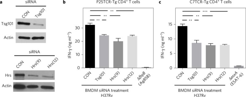
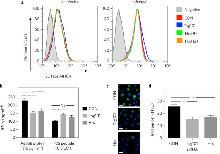
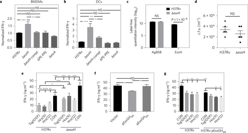
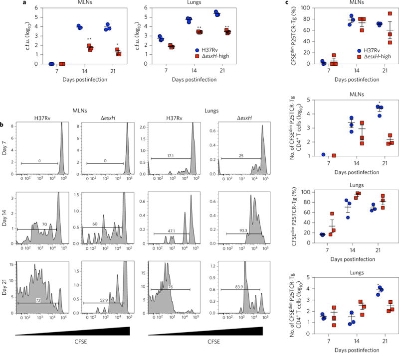
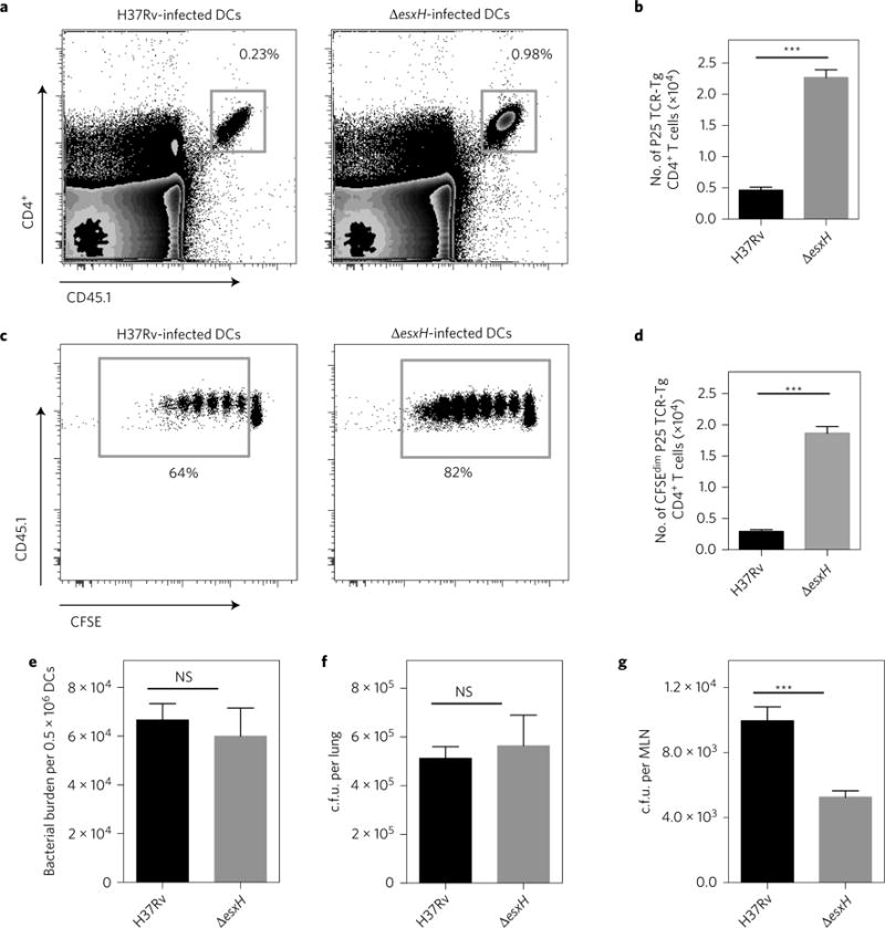
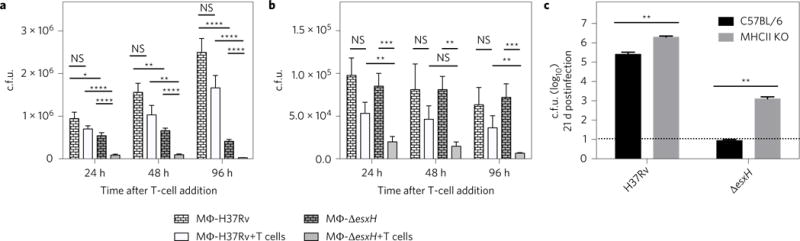
Mycobacterium tuberculosis (Mtb) establishes a persistent infection, despite inducing antigen-specific T-cell responses. Although T cells arrive at the site of infection, they do not provide sterilizing immunity. The molecular basis of how Mtb impairs T-cell function is not clear. Mtb has been reported to block major histocompatibility complex class II (MHC-II) antigen presentation; however, no bacterial effector or host-cell target mediating this effect has been identified. We recently found that Mtb EsxH, which is secreted by the Esx-3 type VII secretion system, directly inhibits the endosomal sorting complex required for transport (ESCRT) machinery. Here, we showed that ESCRT is required for optimal antigen processing; correspondingly, overexpression and loss-of-function studies demonstrated that EsxH inhibited the ability of macrophages and dendritic cells to activate Mtb antigen-specific CD4+ T cells. Compared with the wild-type strain, the esxH-deficient strain induced fivefold more antigen-specific CD4+ T-cell proliferation in the mediastinal lymph nodes of mice. We also found that EsxH undermined the ability of effector CD4+ T cells to recognize infected macrophages and clear Mtb. These results provide a molecular explanation for how Mtb impairs the adaptive immune response.


Conflict of interest statement

Competing interests

The authors declare no competing financial interests.

Figures

Figure 1
Figure 1. ESCRT promotes antigen presentation by Mtb-infected macrophages
a, BMDMs were transfected with small interfering RNA (siRNA) pools targeting Tsg101, individual siRNAs targeting Hrs (numbers 9 and 12) or a non-targeting control (CON). After 96 h, the lysates were analysed by western blotting. Actin served as a loading control. Images shown are from one experiment, representative of at least three independent experiments. b,c, BMDMs were treated with siRNAs as indicated and infected 3 d later with Mtb H37Rv (multiplicity of infection of 3–5), ΔfbpB or ΔesxA strain. The following day, infected BMDMs were co-cultured for 24 h with P25TCR-Tg (b) or C7TCR-Tg (c) CD4+ T cells, and IFN-γ was measured from culture supernatants by an enzyme-linked immunosorbent assay (ELISA). Results reflect the mean ± s.e.m. from at least four replicates and are representative of at least three independent experiments. **P = 0.005, ***P = 0.0001 (unpaired Student’s t-test).
Figure 2
Figure 2. ESCRT promotes antigen presentation by facilitating antigen processing
a–d, BMDMs were treated with siRNAs targeting Hrs (numbers 9 and 12 in panel a, number 9 in b–d) or Tsg101 (pool). (a) Mtb-infected and uninfected BMDMs were immunostained with anti-mouse MHC-II (I-A/I-E) and analysed by flow cytometry. Shaded histograms indicate unstained controls. Data are representative of two independent experiments. (b) BMDMs were incubated with recombinant Ag85B protein or peptide P25 and co-cultured with P25TCR-Tg T cells. IFN-γ was measured by ELISA. Results reflect means ± s.e.m. from at least three replicates. (c) BMDMs were treated with DQ-OVA and the level of green fluorescence was determined using fluorescence microscopy. Images presented are representative of three independent experiments. FITC, fluorescein isothiocyanate. Scale bars, 20 μm. d, Graph shows means ± s.e.m. of the mean fluorescence intensity (MFI) of at least 50 cells for each condition. *P < 0.05, **P < 0.01 (unpaired Student’s t-test); NS, not significant.
Figure 3
Figure 3. EsxGHMt impairs antigen presentation
a,b, BMDMs (a) or DCs (b) were infected with Mtb H37Rv, ΔesxH (mc27846), ΔesxH complemented with EsxGH (ΔesxH-compl), Δpe5ppe4 (mc27848; ΔPE-PPE) or ΔesxA, and co-cultured with P25TCR-Tg CD4+ T cells. Results were normalized to H37Rv infection from at least three independent experiments and reflect means ± s.e.m. c, Mass spectrometry and label-free quantitation algorithms were used to determine normalized relative protein levels of Ag85B and EsxH in culture filtrate from the wild type and the ΔesxH mutant. Results are combined from three independent experiments and reflect means ± s.e.m. d, Bacterial burden in H37Rv- or ΔesxH-infected BMDMs at 24 h postinfection. Results are representative of two independent experiments and reflect means ± s.e.m. e, BMDMs were treated with siRNA and infected with either H37Rv or ΔesxH and co-cultured with P25TCR-Tg CD4+ T cells as above. One-way analysis of variance (ANOVA) was used to compare the siRNA control (CON) sample with ESCRT-silenced samples for both H37Rv and ΔesxH infection. For each siRNA, H37Rv was compared with ΔesxH. Results are representative of three independent experiments and reflect means ± s.e.m. All significant results are indicated, f, BMDMs were infected with H37Rv containing empty vector, EsxGHMt or EsxGHMs for 24 h and co-cultured with P25TCR-Tg CD4+ effector T cells. Results are representative of two independent experiments and reflect means ± s.e.m. g, BMDMs were treated with the indicated siRNAs and infected with Mtb containing empty vector or EsxGHMt as indicated and co-cultured with P25TCR-Tg CD4+ T cells. One-way ANOVA was used to compare the siRNA control sample with ESCRT-silenced samples for both vector control and EsxGHMt; the vector control strain was compared with the EsxGHMt strain for each siRNA treatment. Results are representative of three independent experiments and reflect means ± s.e.m. All significant differences are indicated. For a,b,e–g, IFN-γ released into culture supernatants was measured by an ELISA. *P <0.05, **P<0.01, ***P<0.005, ****P <0.0001 (unpaired Student’s t-test); NS, not significant.
Figure 4
Figure 4. Fewer ΔesxH c.f.u. are required to initiate proliferation in the MLNs and lungs
Mice with adoptively transferred CFSE-labelled P25TCR-Tg CD4+ T cells were infected by aerosol with H37Rv or high-dose ΔesxH. a, The number of c.f.u. in the MLNs and lungs was quantified at the indicated times (n = 3 mice per time point for each bacterial strain). Results reflect means ± s.e.m. b, Representative CFSE dilution profiles of adoptively transferred CFSE-labelled P25TCR-Tg CD4+ T cells in the MLNs and lung at different time points after aerosol infection. The y axis indicates the number of events. The horizontal bar indicates the percentage of proliferating cells. c, Quantitation of proliferating P25TCR-Tg CD4+ T cells in the MLNs and lungs after infection with H37Rv or ΔesxH, as determined by flow cytometry. Results reflect means ± s.e.m. Results are representative of two similar experiments. *P< 0.05, **P < 0.01 (unpaired Student’s t-test).
Figure 5
Figure 5. EsxGHMt impairs priming of naive CD4+ T cells in vivo
a, Representative fluorescence-activated cell sorting plots showing the frequency of adoptively transferred P25TCR-Tg CD4+ T cells in the MLNs of MHC-II/ mice 60 h after intratracheal transfer of MHC-II+/+ DCs infected with either H37Rv or ΔesxH. b, Quantitation of total P25TCR-Tg CD4+ T cells in the MLNs of the groups of mice shown in (a). c, Frequency of CFSE-labelled P25TCR-Tg CD4+ T cells undergoing proliferation in the MLNs of mice 60 h after intratracheal transfer of either H37Rv- or ΔesxH-infected MHC-II+/+ DCs. d, Quantitation of P25TCR-Tg CD4+ T cells that have undergone at least one cycle of proliferation (CFSEdim) in MLNs of groups of mice shown in a–c. e, Bacterial burden in H37Rv- or ΔesxH-infected BMDCs used for intratracheal transfer. Data are expressed as means ± s.e.m. of infected BMDCs in three replicates. f,g, Bacterial burden in lungs f or MLNs g of mice 60 h after receiving intratracheal BMDCs infected with either H37Rv or ΔesxH. All data are from one experiment expressed as mean ± s.e.m. of three pools of mice (n = 6) per experimental group. ***P < 0.001 (Student’s t-test); NS, not significant.
Figure 6
Figure 6. EsxH protects Mtb from CD4+ T-cell mediated killing
a, BMDMs infected with H37Rv (wild type) or ΔesxH for 24 h were cultured for an additional 96 h in the absence or presence of P25TCR-Tg CD4+ cells as indicated, and the number of c.f.u. was determined. b, BMDMs treated with IFN-γ (activated BMDMs) the day before they were infected with H37Rv (wild type) or ΔesxH were incubated at 24 h postinfection with or without P25TCR-Tg CD4+ cells, and the number of c.f.u. was determined at the indicated time points. Data in a and b are expressed as means ± s.e.m. from two independent experiments with at least eight individual replicates per experimental group. *P<0.05, **P<0.01, ***P<0.001, ****P<0.0001 (Student’s t-test corrected for multiple comparisons using the Holm-Sidak method). c, C57BL/6 and MHC-II KO mice were infected with H37Rv or ΔesxH by aerosol (102 c.f.u. for both strains), and the number of c.f.u. was determined in the lungs at 21 d postinfection. Dashed horizontal line indicates the limit of detection. Data are from one experiment expressed as means ± s.e.m. (n = 5 per group). **P <0.01 (unpaired Student’s t-test). MΦ, macrophage; KO, knockout.

Comment in

References

    1. Wolf AJ, et al. Initiation of the adaptive immune response to Mycobacterium tuberculosis depends on antigen production in the local lymph node, not the lungs. J Exp Med. 2008;205:105–115. - PMC - PubMed
    1. MacMicking JD. Interferon-inducible effector mechanisms in cell-autonomous immunity. Nat Rev Immunol. 2012;12:367–382. - PMC - PubMed
    1. Fortune SM, et al. Mycobacterium tuberculosis inhibits macrophage responses to IFN-γ through myeloid differentiation factor 88-dependent and -independent mechanisms. J Immunol. 2004;172:6272–6280. - PubMed
    1. Noss EH, et al. Toll-like receptor 2-dependent inhibition of macrophage class II MHC expression and antigen processing by 19-kDa lipoprotein of Mycobacterium tuberculosis. J Immunol. 2001;167:910–918. - PubMed
    1. Pennini ME, Pai RK, Schultz DC, Boom WH, Harding CV. Mycobacterium tuberculosis 19-kDa lipoprotein inhibits IFN-γ-induced chromatin remodeling of MHC2TA by TLR2 and MAPK signaling. J Immunol. 2006;176:4323–4330. - PubMed

MeSH terms

Substances