. 2016 Dec 5:2:16250.
doi: 10.1038/nmicrobiol.2016.250.

Induction and suppression of antiviral RNA interference by influenza A virus in mammalian cells

Affiliations

Induction and suppression of antiviral RNA interference by influenza A virus in mammalian cells

Yang Li et al. Nat Microbiol. .

Abstract

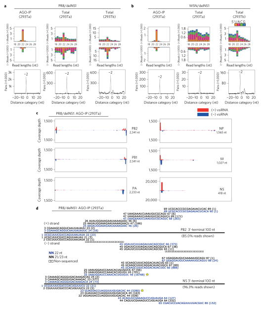
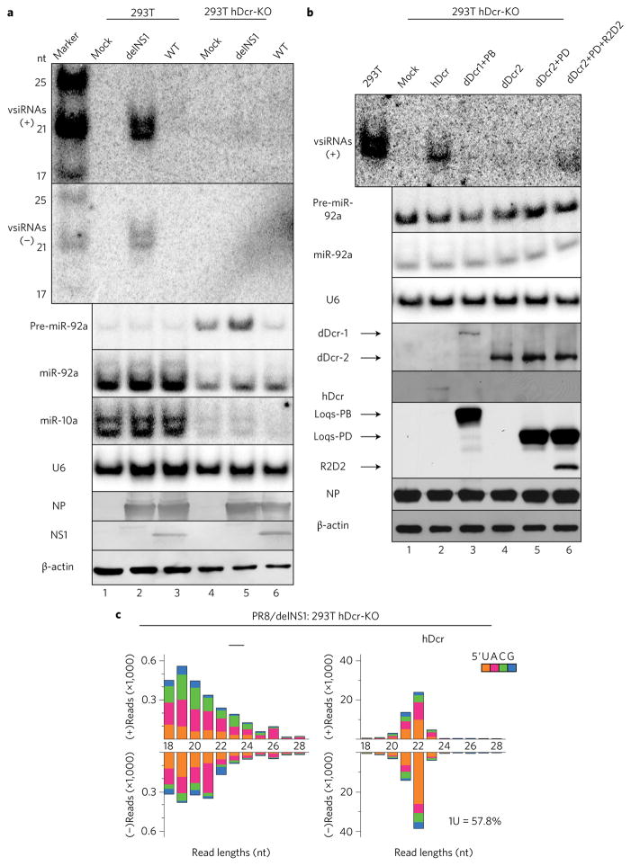
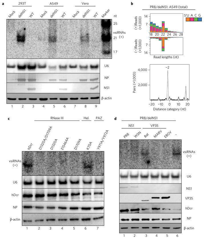
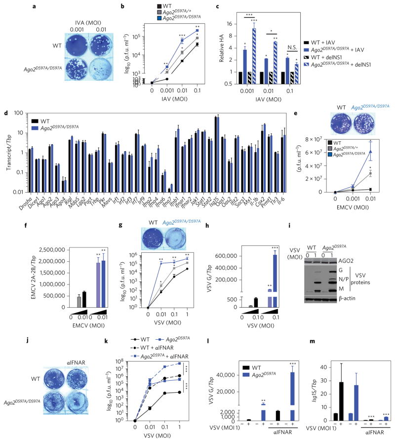
Influenza A virus (IAV) causes annual epidemics and occasional pandemics, and is one of the best-characterized human RNA viral pathogens1. However, a physiologically relevant role for the RNA interference (RNAi) suppressor activity of the IAV non-structural protein 1 (NS1), reported over a decade ago2, remains unknown3. Plant and insect viruses have evolved diverse virulence proteins to suppress RNAi as their hosts produce virus-derived small interfering RNAs (siRNAs) that direct specific antiviral defence4-7 by an RNAi mechanism dependent on the slicing activity of Argonaute proteins (AGOs)8,9. Recent studies have documented induction and suppression of antiviral RNAi in mouse embryonic stem cells and suckling mice10,11. However, it is still under debate whether infection by IAV or any other RNA virus that infects humans induces and/or suppresses antiviral RNAi in mature mammalian somatic cells12-21. Here, we demonstrate that mature human somatic cells produce abundant virus-derived siRNAs co-immunoprecipitated with AGOs in response to IAV infection. We show that the biogenesis of viral siRNAs from IAV double-stranded RNA (dsRNA) precursors in infected cells is mediated by wild-type human Dicer and potently suppressed by both NS1 of IAV as well as virion protein 35 (VP35) of Ebola and Marburg filoviruses. We further demonstrate that the slicing catalytic activity of AGO2 inhibits IAV and other RNA viruses in mature mammalian cells, in an interferon-independent fashion. Altogether, our work shows that IAV infection induces and suppresses antiviral RNAi in differentiated mammalian somatic cells.


Conflict of interest statement

Competing interests

The authors declare no competing financial interests.

Figures

Figure 1
Figure 1. Production of viral siRNAs in mature human somatic cells
a,b, Size distribution and abundance (per million total mature miRNAs) of total virus-derived small RNAs (vsRNAs) sequenced either directly from two human 293T cell lines (293Ta and 293Tb) 24 h after infection with delNS1 mutants of PR8 and WSN strains (total), or after AGO co-IP from the infected 293Ta cells (AGO-IP). Strong enrichment of total and AGO-bound 22 nt RNAs of both PR8/delNS1 and WSN/delNS1 for pairs (−2 peak) of canonical vsiRNAs with 2 nt 3′ overhangs was detected by computing total pairs of 22 nt vsRNAs with different lengths of base-pairing. The 5′-terminal nt of vsRNAs is indicated by colour. c, Relative abundance of 21–23 nt vsiRNA hotspots mapped to PR8/delNS1 genomic RNAs, presented from the 3′ end (left) to the 5′ end (right). The genome segments haemagglutinin (HA) and NA, which are targeted by an extremely low density of vsiRNAs, are shown in Supplementary Fig. 1. d, Read sequences along the 3′-terminal 100 nt of PR8/delNS1 mutant genome segments PB2 and NS. Read counts (in brackets), read length, non-sequenced reads, genomic position and percentage of total reads mapped to the region are indicated. The RNAs complementary to the positive (+) or negative (−)-strand vsiRNAs marked by a star were used subsequently as the probes for northern detection of the influenza vsiRNAs.
Figure 2
Figure 2. Wild-type (WT) hDicer is necessary and sufficient for the biogenesis of human vsiRNAs in differentiated somatic cells
a, Northern detection of the influenza vsiRNAs in WT and hDicer-knockout (hDCr-KO) 293T cells 24 h after infection with the WT or mutant (delNS1) viruses of the PR8 strain. b, Production of influenza vsiRNAs in PR8/delNS1-infected hDcr-KO 293T cells ectopically expressing hDcr, dDicer-2 (dDcr2), dDcr1+Loquacious (Loqs) isoform-PB (PB), dDcr2+Loqs-PD (PD) or dDcr2+PD+R2D2, as indicated. The same sets of RNA and protein samples were used for northern or western blot detection of the positive (+) or negative (−)-strand influenza vsiRNAs (RNA sequences marked by a star in Fig. 1d), precursor microRNA-92a (pre-miR-92a), miR-92a, miR-10a, U6 RNA, viral NP and NS1 proteins, hDcr, the tagged Drosophila proteins or β-actin. Each experiment was repeated twice, with reproducible results. c, Properties of the influenza vsiRNAs (per million total reads) sequenced from PR8/delNS1-infected hDcr-KO 293T cells ectopically expressing hDcr (right). The 5′-terminal nt of the vsiRNAs is indicated by colour and the percentage of 1U vsiRNAs is shown. The sRNAs mapped to the viral genome in PR8/delNS1-infected hDcr-KO 293T cells were low in abundance and exhibited a random size distribution (left).
Figure 3
Figure 3. Induction and suppression of influenza vsiRNA biogenesis in distinct human and monkey somatic cells
a, Northern detection of the influenza vsiRNAs in human (293T and A549) and monkey (Vero) cells 24 h after infection with WT or mutant (delNS1) viruses of the PR8 strain. b, Properties of influenza vsiRNAs (per million total mature miRNAs) sequenced directly without AGO co-IP from PR8/delNS1-infected A549 cells. c, Production of influenza vsiRNAs in PR8/delNS1-infected hDcr-KO 293T cells ectopically expressing WT hDcr, or hDcr mutants carrying the point amino acid mutations known to disrupt the function of the helicase (Hel.; K70A), PAZ (Y971A and Y972A), RNase IIIA (D1320A and E1564A) or RNase IIIB (D1709A) domain of hDcr. d, Suppression of influenza vsiRNA biogenesis in PR8/delNS1-infected hDcr-KO 293T cells ectopically expressing hDcr by NS1 of IAV strain PR8 or WSN, FLAG-tagged VP35 of Ebola virus (EBOV), Marburg virus (MARV) or bat, as indicated. The same sets of RNA and protein samples were used for northern or western blot detection of the positive (+)-strand influenza vsiRNAs, U6 RNA, viral NP and NS1 proteins, FLAG-tagged VP35 variants, hDcr or β-actin as in Fig. 2. Each experiment was repeated twice, with reproducible results.
Figure 4
Figure 4. AGO2 slicing activity restricts IAV, EMCV and VSV in mammalian somatic cells
a,b, Representative plaque assay image of IAV (PR8)-infected (indicated MOIs for 16 h) MEFs from WT mice (black) or mice carrying one (Ago2D597A/+, grey) or two (Ago2D597A/D597A, blue) alleles of catalytic-inactive AGO2. Virus titres quantified as plaque-forming units (p.f.u.) per ml for the indicated MOIs are shown. c, Relative influenza HA RNA as quantified by qPCR in WT or Ago2D597A/D597A MEFs infected with IAV (PR8) or IAV delNS1 mutant. d, Expression of RNAi components, type I IFNs and IFN-stimulated genes (ISGs) in WT (black) or Ago2D597A/D597A (blue) MEFs following influenza infection. e,f, Virus levels in WT (black), Ago2D597A/+ (grey) and Ago2D597A/D597A (blue) MEFs infected with EMCV at indicated MOIs for 16 h as measured by plaque assays of cell supernatants (e) or qPCR of EMCV 2A-2B RNA relative to the housekeeping gene TATA box protein (Tbp) (f). gi, Virus levels in WT (black), Ago2D597A/+ (grey) and Ago2D597A/D597A (blue) MEFs infected with VSV, at the indicated MOIs, for 16 h as measured by plaque assays of cell supernatants (g) or qPCR of VSV-G RNA relative to Tbp (h) or immunoblot of VSV G, N, P and M proteins (i). jl, Virus levels in WT (black) and Ago2D597A/D597A (blue) MEFs infected with VSV in the presence of 10 μg ml−1 anti-IFNAR antibody (aIFNAR; MAR1-5A3, dotted lines) as quantified by plaque assays (j,k) or qPCR of VSV-G RNA (l). m, Expression of IFN-stimulated gene 15 (Isg15) relative to Tbp as measured by qPCR-confirmed IFNAR blocking. All data are two separate lines of MEFs performed two to three independent times in triplicate, combined. All error bars are s.e.m. *P < 0.05, **P < 0.01, ***P < 0.001, measured by an unpaired t-test or two-way ANOVA (Prism). N.S., not significant.

Comment in

References

    1. Medina RA, García-Sastre A. Influenza A viruses: new research developments. Nat Rev Microbiol. 2011;9:590–603. - PMC - PubMed
    1. Li WX, et al. Interferon antagonist proteins of influenza and vaccinia viruses are suppressors of RNA silencing. Proc Natl Acad Sci USA. 2004;101:1350–1355. - PMC - PubMed
    1. Marc D. Influenza virus non-structural protein NS1: interferon antagonism and beyond. J Gen Virol. 2014;95:2594–2611. - PubMed
    1. Lindenbach BD, Rice CM. RNAi targeting an animal virus: news from the front. Mol Cell. 2002;9:925–927. - PubMed
    1. Haasnoot J, Westerhout EM, Berkhout B. RNA interference against viruses: strike and counterstrike. Nat Biotechnol. 2007;25:1435–1443. - PMC - PubMed