. 2017 Jan;23(1):128-135.
doi: 10.1038/nm.4244. Epub 2016 Dec 5.

Interleukin-33-induced expression of PIBF1 by decidual B cells protects against preterm labor

Affiliations

Interleukin-33-induced expression of PIBF1 by decidual B cells protects against preterm labor

Bihui Huang et al. Nat Med. 2017 Jan.

Abstract

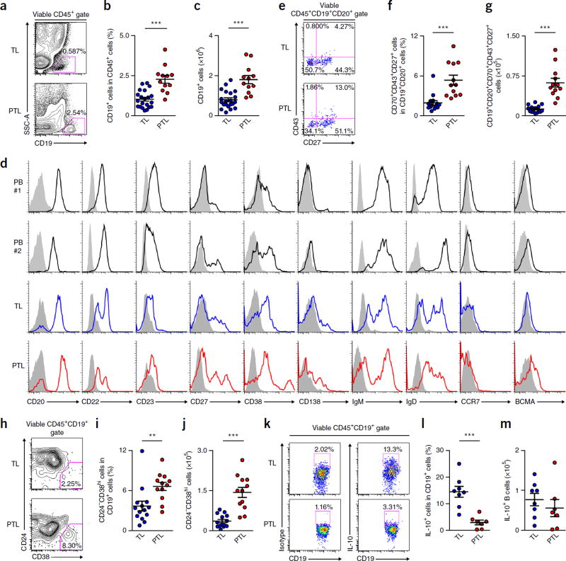
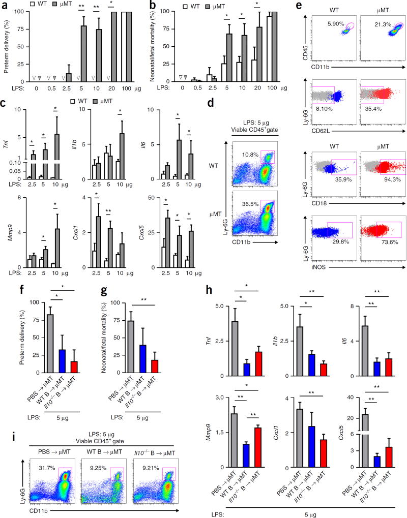
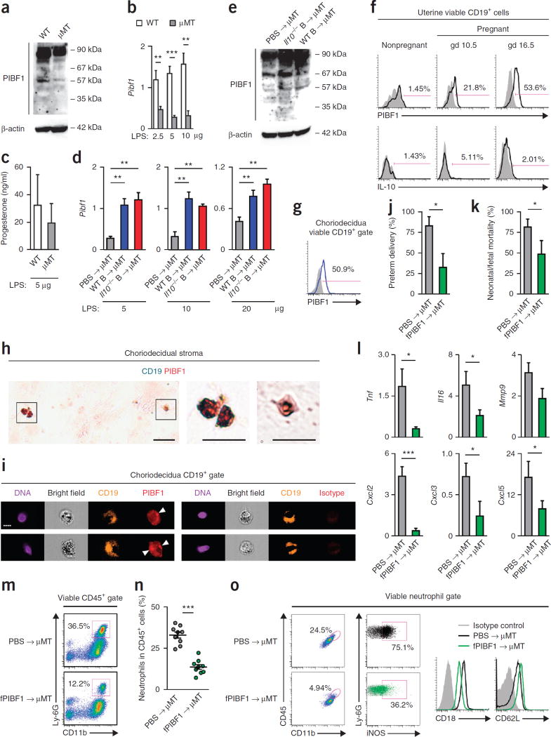
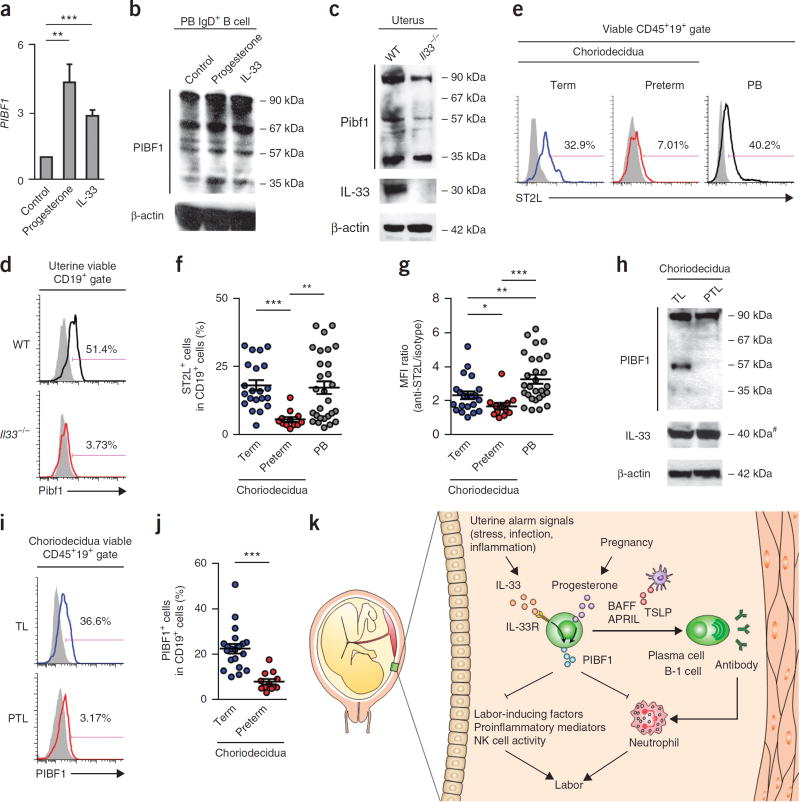
Preterm birth (PTB) is a leading cause of neonatal death worldwide. Intrauterine and systemic infection and inflammation cause 30-40% of spontaneous preterm labor (PTL), which precedes PTB. Although antibody production is a major immune defense mechanism against infection, and B cell dysfunction has been implicated in pregnancy complications associated with PTL, the functions of B cells in pregnancy are not well known. We found that choriodecidua of women undergoing spontaneous PTL harbored functionally altered B cell populations. B cell-deficient mice were markedly more susceptible than wild-type (WT) mice to PTL after inflammation, but B cells conferred interleukin (IL)-10-independent protection against PTL. B cell deficiency in mice resulted in a lower uterine level of active progesterone-induced blocking factor 1 (PIBF1), and therapeutic administration of PIBF1 mitigated PTL and uterine inflammation in B cell-deficient mice. B cells are a significant producer of PIBF1 in human choriodecidua and mouse uterus in late gestation. PIBF1 expression by B cells is induced by the mucosal alarmin IL-33 (ref. 9). Human PTL was associated with diminished expression of the α-chain of IL-33 receptor on choriodecidual B cells and a lower level of active PIBF1 in late gestation choriodecidua. These results define a vital regulatory cascade involving IL-33, decidual B cells and PIBF1 in safeguarding term pregnancy and suggest new therapeutic approaches based on IL-33 and PIBF1 to prevent human PTL.


Conflict of interest statement

COMPETING FINANCIAL INTERESTS

The authors declare no competing financial interests.

Figures

Figure 1
Figure 1
Human choriodecidua harbors B cells that are dysregulated in PTL. (a,b) Frequency of CD19+ B cells in CD45+ cells in choriodecidua of women with TL (n = 21) or PTL (n = 12). (c) Calculated numbers of CD19+ B cells recovered from choriodecidual tissues of women with TL (n = 21) or PTL (n = 12). (d) Expression of CD20, CD22, CD23, CD27, CD38, CD138, IgM, IgD, CCR7 and BCMA on CD19+ B cells in peripheral blood (PB) of two nonpregnant healthy donors (black histogram) and choriodecidual tissues of women with TL (blue histogram) or PTL (red histogram). Shaded histograms indicate the staining of CD19+ B cells in the respective tissues using isotype control antibodies. The results summarize the profiles of 20 healthy blood donors, 16 women with TL and 12 women with PTL. (e,f) CD43 and CD27 expression on CD19+CD20+CD70 B cells and the percentage of CD19+CD20+CD70CD43+CD27+ cells in CD19+CD20+ B cells in choriodecidual tissues of women with TL (n = 15) or PTL (n = 12). (g) Calculated total numbers of CD19+CD20+CD70CD43+CD27+ cells recovered from choriodecidual tissues of women with TL (n = 15) or PTL (n = 12). (h,i) CD24 and CD38 expression on CD19+ B cells and the frequency of CD24CD38hi PCs in CD19+ B cells in choriodecidual tissues of women with TL (n = 14) or PTL (n = 12). (j) Calculated total numbers of CD24CD38hi PCs recovered from choriodecidual tissues of women with TL (n = 14) or PTL (n = 12). (k,l) Frequency of IL-10+ B cells in CD19+ B cells in choriodecidual tissues of women with spontaneous TL (n = 8) or spontaneous PTL (n = 7). (m) Calculated total numbers of IL-10+ B cells recovered from choriodecidual tissues of women with TL (n = 8) or PTL (n = 7). **P < 0.01, ***P < 0.001, by two-tailed t test (b,c,g,i,j,l,m) or two-tailed Mann–Whitney U test (f,i).
Figure 2
Figure 2
B cells confer resistance to inflammation-associated PTL independently of IL-10. (a,b) Rates of preterm delivery and neonatal/fetal mortality of WT or µMT mice 24 h after receiving LPS administered on gd 16.5. Arrowheads indicate no PTL or neonatal/fetal mortality. (c) Fold change of Tnf, Il1b, Il6, Mmp9, Cxcl1 and Cxcl5 transcripts in uterine tissues of WT or µMT mice 24 h after receiving LPS, relative to the gene transcripts in uterine tissues of the respective strain of mice that did not receive LPS. (d) Frequency of CD11b+Ly-6G+ neutrophils in CD45+ cells in uterine tissues of representative WT and µMT mice 24 h after receiving 5 µg LPS. (e) Expression of surface CD11b, CD18, CD62L and intracellular iNOS by viable neutrophils in uterine tissues of a WT or µMT mouse 24 h after receiving 5 µg LPS. (f,g) Rate of preterm delivery and neonatal/fetal mortality on gd 17.5 of µMT mice that received either PBS or WT or Il10−/− B cells on gd 14.5 and 5 µg LPS on gd 16.5. (h) Fold change of Tnf, Il1b, Il6, Mmp9, Cxcl1 and Cxcl5 transcripts in uterine tissues of gd 17.5 µMT mice after receiving either PBS or WT or Il10−/− B cells on gd 14.5 and 5 µg LPS, relative to the gene transcripts in uterine tissues of the respective mice that did not receive LPS. (i) Frequency of CD11b+Ly-6G+ neutrophils in CD45+ cells in uterine tissues of gd 17.5 µMT mice after receiving either PBS or WT or Il10−/− B cells on gd 14.5 and 5 µg LPS on gd 16.5. (j) Fold change of Il10, Tgfb1 and Ebi3 transcripts in uterine tissues of WT or µMT mice 24 h after receiving the indicated dose of LPS, relative to the gene transcripts in uterine tissues of the respective mice that did not receive LPS. Data in a and b represent the results of six WT mice (PBS, 0.5 and 10 µg LPS groups), seven WT mice (2.5 and 5 µg LPS groups), four WT mice (20 µg LPS group), five µMT mice (PBS and 20 µg LPS groups), eight µMT mice (2.5 and 10 µg LPS groups), nine µMT mice (0.5 µg LPS group), ten µMT mice (5 µg LPS group) or four WT or µMT mice (100 µg LPS group) per group. Data in c represent the results of five mice per group. Data in d and e represent the results of seven WT mice and nine µMT mice. Data in f–i represent the results of five mice per group. *P < 0.05, **P < 0.01, by Fisher’s exact test (a,f), one-tailed Mann–Whitney U test (b,g) or one-tailed t test (c,h).
Figure 3
Figure 3
B cells protect against PTL via PIBF1-dependent suppression of uterine inflammation. (a) Western blot analysis (n = 3) of PIBF1 in uterine tissues of WT or µMT mice on gd 16.5. (b) Fold change of Pibf1 transcript in uterine tissues of WT or µMT mice 24 h after receiving LPS given on gd 16.5, relative to the gene transcripts in uterine tissues of the respective mice that did not receive LPS. (c) ELISA of serum progesterone concentrations in WT and µMT mice 24 h after 5 µg LPS challenge. (d) Fold change of Pibf1 transcript in uterine tissues of gd 17.5 µMT mice after receiving either PBS or WT or Il10−/− B cells on gd 14.5 and 5 µg LPS on gd 16.5, relative to the gene transcripts in uterine tissues of the respective mice that did not receive LPS. (e) Western blot analysis (n = 3) of PIBF1 in uterine tissues of µMT mice on gd 16.5 that received WT or Il10−/− B cells on gd 14.5. (f) Flow cytometry analysis of Pibf1 and IL-10 expression by uterine CD19+ B cells in nonpregnant WT mice and pregnant WT mice on gd 10.5 or 16.5. (g) Flow cytometric analysis of PIBF1 expression by choriodecidual CD19+ B cells in a woman with TL. (h) Immunohistochemical analysis of TL choriodecidual stroma for CD19 (turquoise) and PIBF1 (red). Scale bars, 20 µm. (i) Imaging flow cytometry analysis of PIBF1 expression by TL choriodecidual CD19+ B cells. Arrowheads point to concentrated perinuclear PIBF1 staining. Scale bar, 7 µm. (j,k) Rates of preterm delivery and neonatal/fetal mortality on gd 17.5 of µMT mice that received either PBS or fPIBF1 and 5 µg LPS on gd 16.5. (l) Fold change of Tnf, Il6, Mmp9, Cxcl2, Cxcl3 and Cxcl5 transcripts in uterine tissues of µMT mice 24 h after PBS or fPIBF1 administration and 5 µg LPS, relative to the gene transcripts in uterine tissues of the mice that received PIBF1 but not LPS. (m,n) Frequency of CD11b+Ly-6G+ neutrophils in CD45+ cells in uterine tissues of gd 17.5 µMT mice after receiving either PBS or fPIBF1 and LPS on gd 16.5. (o) Expression of surface CD11b, CD18, CD62L and intracellular iNOS by viable neutrophils in uterine tissues of a µMT mouse 24 h after receiving PBS or fPIBF1 and LPS. Data represent the results from five (a–f) or nine mice (j–o) per group. *P < 0.05, **P < 0.01, ***P < 0.001, by Fisher’s exact test (j), one-tailed Mann–Whitney U test (k) or one-tailed t test (b,d,l,n).
Figure 4
Figure 4
IL-33-dependent PIBF1 expression by decidual B cells is defective in human PTL. (a) Fold change of PIBF1 transcript in human PB IgD+ B cells after 3 d of stimulation with progesterone or IL-33, relative to unstimulated IgD+ B cells. (b) Western blot analysis (n = 4) of PIBF1 in human PB IgD+ B cells after 3 d of treatment with medium (control), progesterone or IL-33. Data in a and b represent the results of nine donors. (c,d) Western blot (n = 3) and flow cytometric analyses of PIBF1 expression in uterine tissue or uterine B cells of age-matched pregnant WT and Il33−/− mice on gd 16.5. Data represent three mice per group. (e) Flow cytometry of surface ST2L on B cells in choriodecidual tissues of a woman with TL or a woman with PTL or peripheral blood of a healthy donor. (f,g) Frequency of ST2L+ B cells and mean fluorescence intensity (MFI) of surface ST2L staining in choriodecidual B cells of women with TL (n = 21) or PTL (n = 12) and peripheral blood B cells of healthy donors (n = 28). (h) Western blot analysis (n = 3) of PIBF1 and IL-33 expression in choriodecidual tissue of a subject with TL and a subject with PTL. Data represent four TL and four PTL subjects. Pound sign (#) indicates IL-33 runs at a molecular weight higher than its predicted molecular weight. (i) Flow cytometric analysis of PIBF1 expression in choriodecidual CD19+ B cells of a woman with TL and a woman with PTL. (j) Frequency of PIBF1+ choriodecidual B cells from TL (n = 20) and PTL (n = 12) subjects. (k) A proposed model of choriodecidual-B-cell-mediated protection against PTL via IL-33-induced expression of PIBF1. B cell migration to choriodecidua in pregnancy may be mediated by signals involving α4 and β7 integrins. In response to choriodecidual IL-33, an alarmin that is released following uterine stress, inflammation and infection, choriodecidual B cells produce PIBF1 to suppress premature parturition, likely via the inhibition of the production of labor-inducing factors, natural killer (NK) cell activity, neutrophil infiltration and activation, and the production of proinflammatory mediators. Thus, IL-33, choriodecidual B cells and PIBF1 constitute a protective axis to promote term pregnancy by counteracting uterine stress, inflammation and infection. In PTL, choriodecidual B cells undergo aberrant expansion, increased activation and differentiation into plasma cells or B-1 cells, and secrete antibodies that can trigger labor-inducing inflammatory cascades, such as complement and neutrophil activation. Furthermore, the protective axis involving IL-33, choriodecidual B cells and PIBF1 is defective in PTL, likely as a result of the aberrant downregulation of ST2L on choriodecidual B cells. *P < 0.05, **P < 0.01, ***P < 0.001, by two-tailed t test (a,j) or two-tailed Mann–Whitney U test (f,g).

Comment in

References

    1. Goldenberg RL, Culhane JF, Iams JD, Romero R. Epidemiology and causes of preterm birth. Lancet. 2008;371:75–84. - PMC - PubMed
    1. Romero R, et al. The preterm parturition syndrome. BJOG. 2006;113(Suppl. 3):17–42. - PMC - PubMed
    1. Jensen F, et al. CD19+CD5+ cells as indicators of preeclampsia. Hypertension. 2012;59:861–868. - PubMed
    1. Zhou CC, et al. Angiotensin receptor agonistic autoantibodies induce pre-eclampsia in pregnant mice. Nat. Med. 2008;14:855–862. - PMC - PubMed
    1. Matthiesen L, et al. Immunology of preeclampsia. In: Markert UR, editor. Immunology of Pregnancy. Vol. 89. Karger, Basel: 2005. pp. 49–61. - PubMed

Publication types

MeSH terms