. 2016 Dec 5;12(12):e1006441.
doi: 10.1371/journal.pgen.1006441. eCollection 2016 Dec.

The Gene Regulatory Network of Lens Induction Is Wired through Meis-Dependent Shadow Enhancers of Pax6

Affiliations

The Gene Regulatory Network of Lens Induction Is Wired through Meis-Dependent Shadow Enhancers of Pax6

Barbora Antosova et al. PLoS Genet. .

Abstract

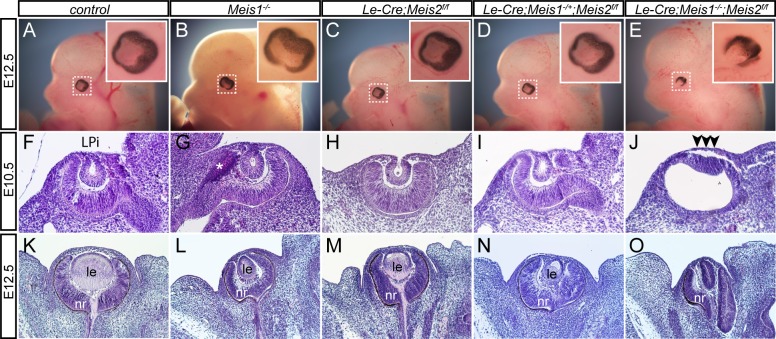
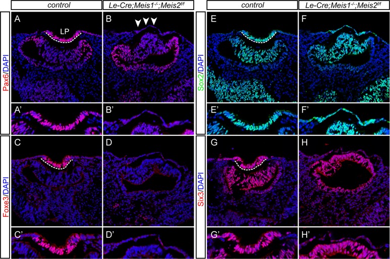
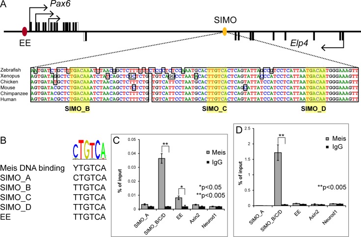
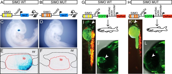
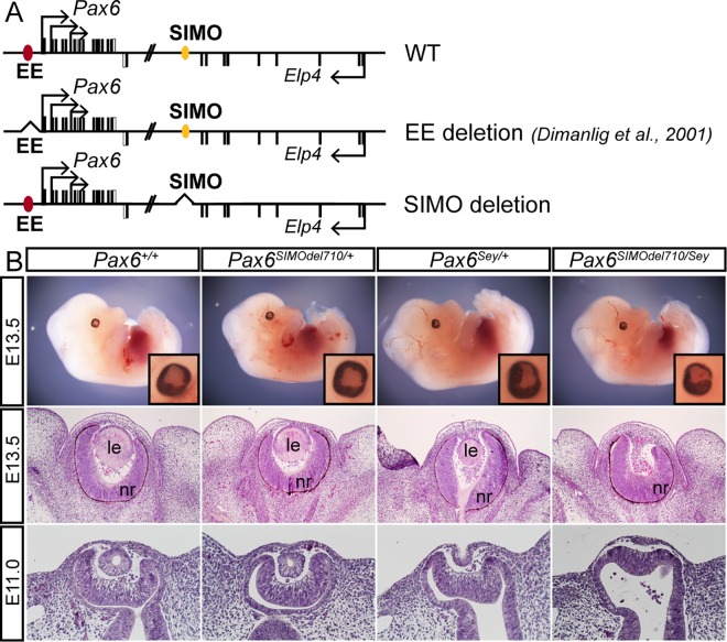
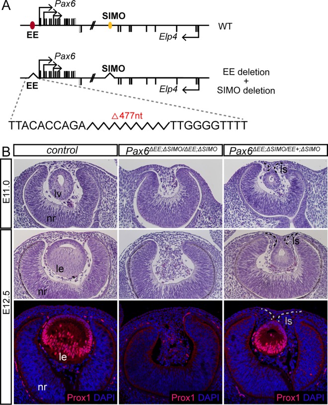
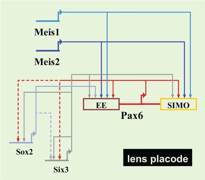
Lens induction is a classical developmental model allowing investigation of cell specification, spatiotemporal control of gene expression, as well as how transcription factors are integrated into highly complex gene regulatory networks (GRNs). Pax6 represents a key node in the gene regulatory network governing mammalian lens induction. Meis1 and Meis2 homeoproteins are considered as essential upstream regulators of Pax6 during lens morphogenesis based on their interaction with the ectoderm enhancer (EE) located upstream of Pax6 transcription start site. Despite this generally accepted regulatory pathway, Meis1-, Meis2- and EE-deficient mice have surprisingly mild eye phenotypes at placodal stage of lens development. Here, we show that simultaneous deletion of Meis1 and Meis2 in presumptive lens ectoderm results in arrested lens development in the pre-placodal stage, and neither lens placode nor lens is formed. We found that in the presumptive lens ectoderm of Meis1/Meis2 deficient embryos Pax6 expression is absent. We demonstrate using chromatin immunoprecipitation (ChIP) that in addition to EE, Meis homeoproteins bind to a remote, ultraconserved SIMO enhancer of Pax6. We further show, using in vivo gene reporter analyses, that the lens-specific activity of SIMO enhancer is dependent on the presence of three Meis binding sites, phylogenetically conserved from man to zebrafish. Genetic ablation of EE and SIMO enhancers demostrates their requirement for lens induction and uncovers an apparent redundancy at early stages of lens development. These findings identify a genetic requirement for Meis1 and Meis2 during the early steps of mammalian eye development. Moreover, they reveal an apparent robustness in the gene regulatory mechanism whereby two independent "shadow enhancers" maintain critical levels of a dosage-sensitive gene, Pax6, during lens induction.


Conflict of interest statement

The authors have declared that no competing interests exist.

Figures

Fig 1
Fig 1. The phenotypic consequences of Meis1 and Meis2 deficiency.
(A-E) At E12.5, external eyes of whole-mount Meis1-/-, Le-Cre;Meis2f/f, Le-Cre;Meis1+/-;Meis2f/f mutant appear comparable to control eye, whereas the eye of Le-Cre;Meis1-/-;Meis2f/f double mutant has abnormal shape. The insets show high magnification of eye region (boxed). (F-O) Hematoxylin-eosin stained parrafin sections show histology of control or mutant E10.5 and E12.5 eyes. (F-H, K-M) Formation of lens placode is followed by invagination of surface ectodem, formation of lens pit (LPi) and subsequent formation of lens in control, Meis1-/- and Le-Cre;Meis2f/f embryos. (I, N) One active Meis1 allele in Le-Cre;Meis1-/+; Meis2f/f embryos is sufficient for lens placode and lens formation. (J, O) In Le-Cre;Meis1-/-;Meis2f/f embryos, deficient for both Meis1 and Meis2, lens development is arrested in pre-placodal stage (arrowheads). * Artefact, le-lens, nr-neural retina.
Fig 2
Fig 2. The expression of lens placode-specific transcription factors is disturbed in Meis1/Meis2 double mutants.
(A-H‘) Cryosections from E10.0 control and Le-Cre;Meis1-/-;Meis2f/f embryos stained with antibody as indicated and nuclei counterstained with DAPI. (B, B‘) Pax6 is not detected in lens surface ectoderm of Le-Cre;Meis1-/-;Meis2f/f embryos (arrowheads) and (D, D‘) expression of the lens differentiation gene Foxe3 is not initiated. (F, F‘) Sox2 is detected in PLE of Meis1/Meis2 double mutants, althouth it failed to thicken. (H, H‘) Finally, expression of Six3 is decreased compared to control. Lens placode (LP) is indicated by dashed line. (A‘-H‘) For clearer examination, lens placode or corresponding lens surface ectoderm region is magnified and shown separately.
Fig 3
Fig 3. Meis proteins bind SIMO element of Pax6 in vivo.
(A) Schematic representation of the Pax6 locus, displaying the exons of Pax6 (black boxes, top strand) and adjacent Elp4 gene (black boxes, bottom strand). Ectoderm enhancer (EE) is indicated with red oval; SIMO enhancer is indicated with yellow oval. The detail of the part of the SIMO shows high conservation across the vertebrate species. In SIMO, five putative Meis binding sites were identified with three, SIMO_B, SIMO_C and SIMO_D (indicated with yellow color), clustered in highly conserved part of the SIMO enhancer. (B) The nucleotide composition of selected putative Meis binding sites found in SIMO and their comparison with Meis consensus binding site and previously identified Meis binding site in EE. (C, D) Results of chromatin immunoprecipitation of Meis-bound DNA fragments performed with the mixture of Meis1-specific and Meis2-specific antibody on chromatin prepared from E10.5 whole embryos (C) or αTN4 mouse lens epithelial cells (D) showing clear enrichment on SIMO enhancer. (C, D) Error bars denote SDs, *p and **p versus control using Student's t‐test.
Fig 4
Fig 4. Characterization of SIMO wild-type and mutant enhancer by reporter gene assays in chick and zebrafish.
(A, B) Schematic view of reporter constructs used for in ovo electroporation of chick embryos. Reporter constructs carry wild-type or mutant mouse SIMO element upstream of hsp68 minimal promoter and β-galactosidase open reading frame. In mutant SIMO Meis binding sites were abolished by introduction of specific single-point mutations changing Meis recognition sequence TGACAG/A into TcACAG/A. (C–F) Whole-mount view or histological sections through the eye of β-galactosidase–stained chick embryos of stage HH21-22 electroporated either with (C, E) wild-type or with (D, F) mutant SIMO fragment. Positive X-gal staining correlates with the activity of reporter constructs. Wild-type SIMO fragment supports reporter construct expression in lens but not the mutant SIMO fragment. (G, H) Schematic view of reporter constructs used for transgenesis in zebrafish. Reporter constructs carry wild-type or mutant zebrafish SIMO element upstream of zebrafish gata2a minimal promoter and EGFP open reading frame. In mutant zebrafish SIMO Meis binding sites were abolished by introduction of specific single-point mutations changing Meis recognition sequence TGACAG/A into TcACAG/A. In order to control for transgenesis efficiency in vivo the reporter genes contain a second cassette composed of a cardiac actin promoter driving the expression of a red fluorescent protein (DsRed). EGFP and DsRed transcriptional units are separated by an insulator. (I-L) Wild-type SIMO enhancer activity is detected at 48 hpf (n = 160, 68% EGFP of DsRed positive), (I, J), but not for the mutant construct (n = 36, 89% EGFP negative of DsRed positive) (K, L). LE—lens, NR—neural retina.
Fig 5
Fig 5. Genetic analysis of SIMO deletion in vivo.
(A) Scheme of wild-type Pax6 locus and alleles carrying EE [17] or SIMO deletion (this study). EE is indicated with red oval and SIMO with yellow oval. (B) Phenotypic consequences of SIMO deletion in Pax6eSIMOdel710/Sey compound heterozygote mice. Whole-mount view of E13.5 embryos of the indicated genotype with eye in the inset (top panel). Histological sections through the eye demonstrating the absence of lens at E13.5 (middle panel) and arrested development prior to lens pit stage at E11.0 in Pax6 SIMOdel710/Sey embryos. nr—neural retina, le-lens.
Fig 6
Fig 6. Genetic analysis of the simultaneous deletion of EE and SIMO in vivo.
(A) Scheme of wild type Pax6 locus, and allele carrying simultaneous deletion of EE and SIMO. EE is indicated with red oval and SIMO with yellow oval. The exact borders of EE deletion are specified by nucleotide sequences flanking the deletion. (B) Phenotypic consequences of simultaneous deletion of EE and SIMO in Pax6ΔEE;ΔSIMO/ΔEE;ΔSIMO embryos. Hematoxylin and eosin stained paraffin sections demonstrating the arrested lens development prior to lens pit stage at E11.0 and absence of lens at E12.5 in Pax6ΔEE;ΔSIMO/ΔEE;ΔSIMO embryos. Immunoflurescent staining for lens marker Prox1 is not detected in E12.5 Pax6ΔEE;ΔSIMO/ΔEE;ΔSIMO embryos. Note that a single allele of intact EE in Pax6ΔEE;ΔSIMO/EE+; ΔSIMO embryos is sufficient for lens formation albeit the resulting lens is much smaller compared to control, and lens stalk is apparent. nr—neural retina, lv – lens vesicle, le – lens, ls – lens stalk.
Fig 7
Fig 7. Current model of transcriptional regulatory network operating during mammalian lens induction.
Direct interactions are indicated with solid lines, whereas dashed lines show possible direct interactions inferred from gain- and loss-of-function studies.

References

    1. Cvekl A, Ashery-Padan R. The cellular and molecular mechanisms of vertebrate lens development. Development. 2014;141(23):4432–47. PubMed Central PMCID: PMC4302924. 10.1242/dev.107953 - DOI - PMC - PubMed
    1. Gunhaga L. The lens: a classical model of embryonic induction providing new insights into cell determination in early development. Philosophical transactions of the Royal Society of London Series B, Biological sciences. 2011;366(1568):1193–203. PubMed Central PMCID: PMC3061103. 10.1098/rstb.2010.0175 - DOI - PMC - PubMed
    1. Bailey AP, Bhattacharyya S, Bronner-Fraser M, Streit A. Lens specification is the ground state of all sensory placodes, from which FGF promotes olfactory identity. Developmental cell. 2006;11(4):505–17. 10.1016/j.devcel.2006.08.009 - DOI - PubMed
    1. Hill RE, Favor J, Hogan BL, Ton CC, Saunders GF, Hanson IM, et al. Mouse small eye results from mutations in a paired-like homeobox-containing gene. Nature. 1991;354(6354):522–5. 10.1038/354522a0 - DOI - PubMed
    1. Hogan BL, Horsburgh G, Cohen J, Hetherington CM, Fisher G, Lyon MF. Small eyes (Sey): a homozygous lethal mutation on chromosome 2 which affects the differentiation of both lens and nasal placodes in the mouse. Journal of embryology and experimental morphology. 1986;97:95–110. - PubMed

MeSH terms