

Somatic GNAQ Mutation is Enriched in Brain Endothelial Cells in Sturge-Weber Syndrome
- PMID: 27919468
- PMCID: PMC5303551
- DOI: 10.1016/j.pediatrneurol.2016.10.010
Somatic GNAQ Mutation is Enriched in Brain Endothelial Cells in Sturge-Weber Syndrome
Abstract
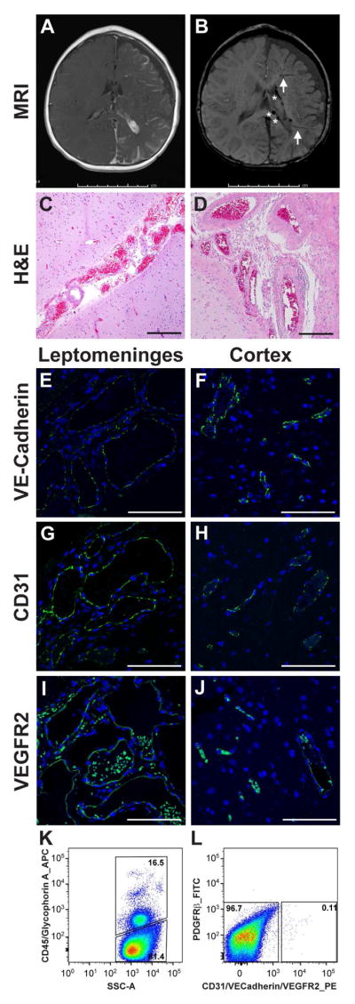
Background: Sturge-Weber syndrome (SWS) is a rare congenital neurocutaneous disorder characterized by facial and extracraniofacial capillary malformations and capillary-venule malformations in the leptomeninges. A somatic mosaic mutation in GNAQ (c.548G>A; p.R183Q) was found in SWS brain and skin capillary malformations. Our laboratory showed endothelial cells in skin capillary malformations are enriched for the GNAQ mutation. The purpose of this study is to determine whether the GNAQ mutation is also enriched in endothelial cells in affected SWS brain.
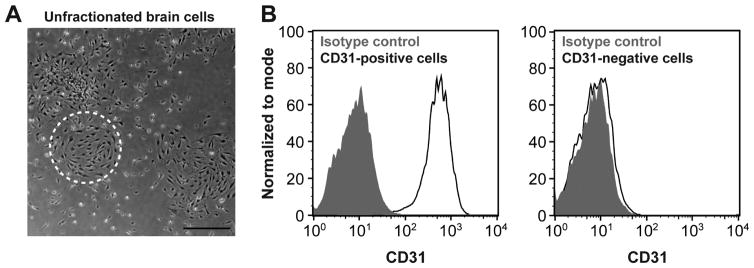
Methods: Two human SWS brain specimens were fractionated by fluorescence-activated cell sorting into hematopoietic (CD45), endothelial (CD31, VE-Cadherin, and vascular endothelial growth factor receptor 2), and perivascular (platelet-derived growth factor receptor beta) cells and cells negative for all markers. The sorted cell populations were analyzed for GNAQ p.R183Q mutation by droplet digital polymerase chain reaction. SWS patient-derived brain endothelial cells were selected by anti-CD31-coated magnetic beads and cultured in endothelial growth medium in vitro.
Results: The GNAQ p.R183Q mutation was present in brain endothelial cells in two SWS specimens, with mutant allelic frequencies of 34.7% and 24.0%. Cells negative for all markers also harbored the GNAQ mutation. The mutant allelic frequencies in these unidentified cells were 9.2% and 8.4%. SWS patient-derived brain endothelial cells with mutant allelic frequencies of 14.7% and 21% survived and proliferated in vitro.
Conclusions: Our study provides evidence that GNAQ p.R183Q mutation is enriched in endothelial cells in SWS brain lesions and thereby reveals endothelial cells as a source of aberrant Gαq signaling. This will help to understand the pathophysiology of SWS, to discover biomarkers for predicting cerebral involvement, and to develop therapeutic targets to prevent neurological impairments in SWS.
Keywords: GNAQ; Sturge–Weber syndrome; capillary malformation; ddPCR; endothelial cell.
Copyright © 2016 Elsevier Inc. All rights reserved.
Conflict of interest statement
of potential conflicts of interest None
Figures
References
-
- Comi AM. Update on Sturge-Weber syndrome: diagnosis, treatment, quantitative measures, and controversies. Lymphat Res Biol. 2007;5:257–264. - PubMed
-
- Sudarsanam A, Ardern-Holmes SL. Sturge-Weber syndrome: from the past to the present. Eur J Paediatr Neurol. 2014;18:257–266. - PubMed
-
- Dutkiewicz AS, Ezzedine K, Mazereeuw-Hautier J, Lacour JP, Barbarot S, Vabres P, Miquel J, Balguerie X, Martin L, Boralevi F, Bessou P, Chateil JF, Leaute-Labreze C. A prospective study of risk for Sturge-Weber syndrome in children with upper facial port-wine stain. J Am Acad Dermatol. 2015;72:473–480. - PubMed
-
- Ch’ng S, Tan ST. Facial port-wine stains - clinical stratification and risks of neuro-ocular involvement. J Plast Reconstr Aesthet Surg. 2008;61:889–893. - PubMed
Publication types
MeSH terms
Substances
Grants and funding
LinkOut - more resources
Full Text Sources
Other Literature Sources
Research Materials
Miscellaneous
