. 2017 Feb:15:127-136.
doi: 10.1016/j.ebiom.2016.11.015. Epub 2016 Dec 1.

Advax, a Delta Inulin Microparticle, Potentiates In-built Adjuvant Property of Co-administered Vaccines

Affiliations

Advax, a Delta Inulin Microparticle, Potentiates In-built Adjuvant Property of Co-administered Vaccines

Masayuki Hayashi et al. EBioMedicine. 2017 Feb.

Abstract

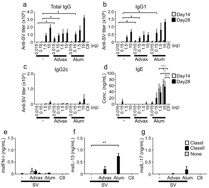
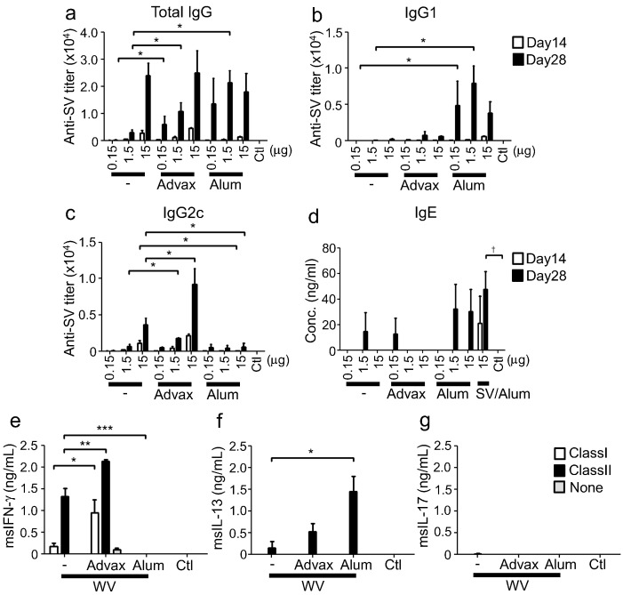
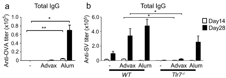
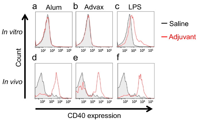
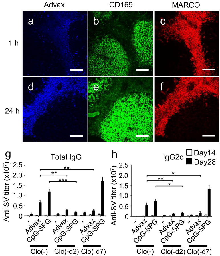
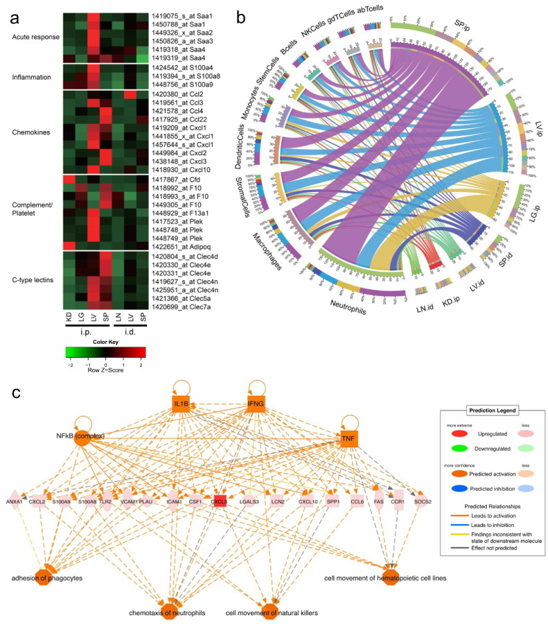
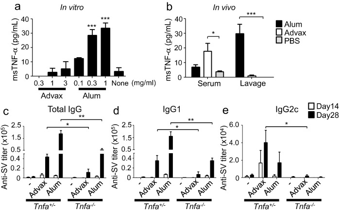
Advax, a delta inulin-derived microparticle, has been developed as an adjuvant for several vaccines. However, its immunological characteristics and potential mechanism of action are yet to be elucidated. Here, we show that Advax behaves as a type-2 adjuvant when combined with influenza split vaccine, a T helper (Th)2-type antigen, but behaves as a type-1 adjuvant when combined with influenza inactivated whole virion (WV), a Th1-type antigen. In addition, an adjuvant effect was not observed when Advax-adjuvanted WV vaccine was used to immunize toll-like receptor (TLR) 7 knockout mice which are unable to respond to RNA contained in WV antigen. Similarly, no adjuvant effect was seen when Advax was combined with endotoxin-free ovalbumin, a neutral Th0-type antigen. An adjuvant effect was also not seen in tumor necrosis factor (TNF)-α knockout mice, and the adjuvant effect required the presences of dendritic cells (DCs) and phagocytic macrophages. Therefore, unlike other adjuvants, Advax potentiates the intrinsic or in-built adjuvant property of co-administered antigens. Hence, Advax is a unique class of adjuvant which can potentiate the intrinsic adjuvant feature of the vaccine antigens through a yet to be determined mechanism.

Keywords: Adjuvant; Inulin; Macrophage; Particle; Vaccine.


Figures

Fig. 1
Fig. 1
Advax induces Th2 responses when combined with a Th2-type antigen. (a-d) On days 0 and 14, C57BL/6J mice (n = 3) were immunized i.m. with SV and adjuvant. Antigen-specific total IgG, IgG1 and IgG2c titers, and total IgE in sera at days 14 and 28 were measured by ELISA. Mice in control (Ctl) group were administered saline. (e-g) On day 28, splenocytes were prepared from mice immunized with 15 μg SV and adjuvant, and stimulated with MHC class I or II epitope peptides of nucleoprotein. After stimulation, IFN-γ, IL-13 and IL-17 in supernatants were measured by ELISA. Results are representatives of three separate experiments. Median and SEM are shown for each group. Statistical significances are indicated, *P < 0.05, **P < 0.01, ***P < 0.001, P < 0.05, †††P < 0.001 by Dunnett's Multiple comparison test.
Fig. 2
Fig. 2
Advax induces Th1 responses when combined with Th1-type antigen. (a–d) on days 0 and 14, C57BL/6J mice (n = 3) were immunized i.m. with WV and adjuvant. 15 μg of SV was immunized with alum on day 0 and 14. Antigen-specific total IgG, IgG1 and IgG2c titres, and total IgE in sera at days 14 and 28 were measured by ELISA. (e–g) On day 28, splenocytes were prepared from mice immunized with 15 μg WV and adjuvant, and stimulated with MHC class I or II specific peptides from nucleoprotein. After stimulation, IFN-γ, IL-13 and IL-17 in supernatants were measured by ELISA. Results are representative of three separate experiments. Median and SEM are shown for each group. Statistical significances are indicated, *P < 0.05, **P < 0.01, ***P < 0.001, P < 0.05 by Dunnett's Multiple comparison test.
Fig. 3
Fig. 3
Advax does not enhance antibody responses to OVA, a Th0-type antigen or to WV in Tlr7−/− mice. (a) C57BL/6J mice (n = 4 or 5) or (b) Tlr7−/− mice (n = 5) were i.m. immunized twice (day 0 and 14) with 100 μg OVA and adjuvant, or 1.5 μg WV alone or together with adjuvant, respectively. Antigen-specific total IgG titers in sera at days 14 and 28 were measured by ELISA. Results are representative of two or three separate experiments. Median and SEM are shown for each group. Statistical significances are indicated, *P < 0.05, **P < 0.01 by Dunnett's Multiple comparison test or student t-test.
Fig. 4
Fig. 4
Advax activates DCs in vivo but not in vitro. (a–c) Bone marrow-derived DCs were stimulated with 1 mg/mL alum, 1 mg/mL Advax or 50 ng/mL LPS for 24 h in vitro, and then the expression of CD40 on pDCs was evaluated. (d–f) C57BL/6J mice were injected with 0.67 mg alum, 1 mg Advax or 50 ng LPS at the base of the tail. Twenty-four hours after injection, the inguinal lymph nodes were collected, treated with DNase I and collagenase. Then, the cells were stained and the expression of CD40 on pDCs was analyzed by FACS. Results are representative of three separate experiments.
Fig. 5
Fig. 5
Macrophages are required for Advax's adjuvant effect. Two-photon microscopy analysis of lymph nodes (a–c) 1 h and (d–f) 24 h after i.d. administration of Brilliant Violet 421-labeled Advax delta inulin particles. CD169+ and MARCO+ macrophages were stained by i.d. injection of anti-CD169-FITC and anti-MARCO-phycoerythrin antibodies 30 min before Advax administration. The pictures were taken by FV1000MPE (Olympus) with 25 × lens (XLPLN25XWMP; Olympus). Scale bar indicates 100 μm. (a, d) Blue indicates Advax, (b, e) green indicates CD169+ macrophages and (c, f) red indicates MARCO+ macrophages. (g, h) Phagocytic cells in lymph nodes were depleted by clodronate liposome injection at the indicated days (-d2 and -d7), and then WV plus Advax was i.d. immunized at d0. Antigen-specific total IgG or IgG2c titers in sera at days 14 and 28 were measured by ELISA. Results are representative of three separate experiments. Median and SEM are shown for each group. Statistical significances are indicated, *P < 0.05, **P < 0.01, ***P < 0.001 by Student's t-test.
Fig. 6
Fig. 6
Advax alters gene expression of IL-1β, CLRs and TNF-α signaling pathways. Whole organ (lung; LG, liver; LV, spleen; SP, kidney; KD, lymph node; LN) transcriptome of 6 h after Advax administration (i.d. or i.p.) alone was obtained by Affimetrix GeneChip (n = 3). (a) Only selected gene probes (FC > 2 and PA = 1) are displayed. (b) Advax-responding cell population analysis was performed (see Materials & Methods; cell population analysis). The left hemispheres of the figure denote the ten immune cell types and right hemispheres denote the samples. The ribbons in the inner circle denote the cell type score of each sample. The colors in the middle layer rings represent either cell types or samples. The outermost ring denotes the percentage of total contribution for every factor (cell type or samples) viewed from their counterpart factor. For example in SP.ip, neutrophils constitute around 30% of the cell population. (c) IPA upstream regulator analysis of Advax-induced genes in the SP was performed. (see Materials & Methods; upstream regulator analysis in IPA). FC: Fold Change, PA: Presence and Absence. (see Materials & Methods; microarray analysis).
Fig. 7
Fig. 7
TNF-α is required for the adjuvant effect of Advax. (a) Peritoneal macrophages were stimulated with Advax or alum for 8 h and TNF-α in supernatants was measured by ELISA. (b) One hour after i.p. injection of 1 mg Advax or 0.67 mg alum, sera and peritoneal lavage fluids were collected and TNF-α levels in the sera and fluids were measured by ELISA. (c–e) On days 0 and 14, heterozygous or Tnf−/− mice (n = 5) were immunized i.m. with 1.5 μg WV and adjuvant. Antigen-specific total IgG, IgG1 and IgG2c titers in sera at days 14 and 28 were measured by ELISA. Results are representative of two separate experiments. Median and SEM are shown for each group. Statistical significances are indicated, *P < 0.05, **P < 0.01, ***P < 0.001 by Dunnett's Multiple comparison test or Student's t-test.

References

    1. Aoshi T., Zinselmeyer B.H., Konjufca V., Lynch J.N., Zhang X., Koide Y., Miller M.J. Bacterial entry to the splenic white pulp initiates antigen presentation to CD8 + T cells. Immunity. 2008;29:476–486. - PubMed
    1. Aoshi T., Carrero J.A., Konjufca V., Koide Y., Unanue E.R., Miller M.J. The cellular niche of Listeria monocytogenes infection changes rapidly in the spleen. Eur. J. Immunol. 2009;39:417–425. - PMC - PubMed
    1. Arase N., Arase H., Park S., Ohno H., Ra C., Saito T. Association with FcRg is essential for activation signal through NKR-P1 (CD161) in natural killer (NK) cells and NK1.1 + T cells. J. Exp. Med. 1997;186:1957–1963. - PMC - PubMed
    1. Coffman R.L., Sher A., Seder R.A. Vaccine adjuvants: putting innate immunity to work. Immunity. 2010;33:492–503. - PMC - PubMed
    1. Cooper P.D., Petrovsky N. Delta inulin: a novel, immunologically active, stable packing structure comprising beta-d-[2 → 1] poly(fructo-furanosyl) alpha-d-glucose polymers. Glycobiology. 2011;21:595–606. - PMC - PubMed

MeSH terms