. 2017 Mar 1:388:64-72.
doi: 10.1016/j.canlet.2016.11.026. Epub 2016 Dec 3.

Modulation of post-translational modifications in β-catenin and LRP6 inhibits Wnt signaling pathway in pancreatic cancer

Affiliations

Modulation of post-translational modifications in β-catenin and LRP6 inhibits Wnt signaling pathway in pancreatic cancer

Bharti Garg et al. Cancer Lett. .

Abstract

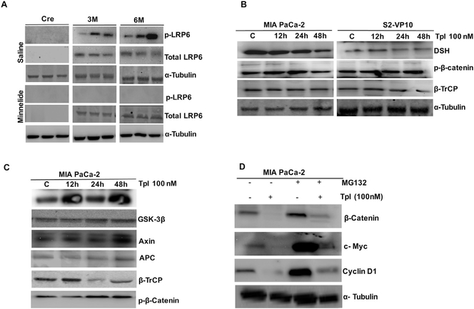
β-Catenin/Wnt signaling pathway is critically regulated in a normal cell by a number of post-translational modifications. In pancreatic cancer however, aberrant activation of this pathway plays a significant role in tumor progression and metastasis. Though a number of studies have focused on understanding Wnt signaling pathway in pancreatic cancer, there has been no systematic study to evaluate molecules that may be affecting this pathway. In the current study, we used a diterpene triepoxide, triptolide, to inhibit post-translational modifications in Wnt pathway and evaluated how this compound may be affecting the intricate signaling that regulates cell proliferation in pancreatic cancer. Our results showed that triptolide inhibits the activation of WNT1, FZD1, and disheveled (DSH) in pancreatic cancer cell lines MIA PaCa-2 and S2-VP10 by inhibiting the phosphorylation of LRP6 and simultaneously blocked translocation of β-catenin to the nucleus by inhibiting its glycosylation. Additionally, inhibition of post-translational modification of the Wnt-signaling pathway also demonstrated regression of tumor growth in a Syngenic Tumor Implantation Model (STIM). Interestingly, these findings suggest Wnt signaling is a vital molecular pathway in pancreatic cancer and may be amenable to targeted drug therapy.

Keywords: OGT; Pancreatic tumor; STIM model; TCF/LEF1; Wnt; β-Catenin.


Conflict of interest statement

Disclosures

The University of Minnesota has a patent for Minnelide™ (which has been licensed to Minneamrita Therapeutics LLC, Moline, IL). AS has ownership interests (including patents) and is a consultant/advisory board member for Minneamrita Therapeutics LLC. SB is a consultant for Minneamrita Therapeutics. This relationship is managed by University of Miami according to its conflict of interest policy. The other authors have nothing to disclose.

Figures

Fig. 1.
Fig. 1.
Evidence of activation of WNT/β-catenin pathway in human and mice model of pancreatic cancer. (A) q-RT PCR analysis of Wnt pathway genes in pancreatic cancer cell lines. (B) Nuclear transport of β-catenin in various human pancreatic cancer cell lines along with HPDEC. (C) Expressional dynamics of Wnt pathway genes during onset and progression of pancreatic cancer in KPC mice (cre, three and six months old) by q-RT PCR. (D) Immunoblotting of β-catenin in cytoplasmic and nuclear protein fractions during the onset and progression of tumor as compared with control cre mice. (E) Immunofluorescence showing the β-catenin expression during the progression of pancreatic tumor. Experiments were performed n = 3 and similar results were obtained each time.
Fig. 2.
Fig. 2.
Effect of inhibition of LRP6 and FZD1 on β-catenin-TCF signaling. (A) Transcriptional changes in β-catenin after treating MIA PaCa-2 and S2-VP10 cell lines with SiLRP6, SiFZD1 and SiLRP6+SiFZD1 for 48 h and overexpression of recombinant rLRP6 and rFZD1 plasmids for 24 h. (B) MIA PaCa-2 and S2-VP10 cytoplasmic and nuclear fractions were isolated using cytoplasmic and nuclear extraction kit. Different fractions of MIA PaCa and S2-VP10 cells were immunoblotted and probed with β-catenin antibody. (C) MIA PaCa-2 and S2-VP10 cells were transfected with TCF/LEF reporter plasmid for 18 h and treated with SiLRP6, SiFZD1 and SiLRP6+SiFZD1 for 48 h, inhibition of LRP6 and FZD were recovered using rLRP6 and rFZD1 plasmids for 24 h, luciferase activity was counted with Promega kit. (D) Expression of Cyclin D1 and c-Myc by q-RT PCR upon LRP6 and FZD silencing in MIA PaCa-2 and S2-VP10 (Supplementary Fig. 4) cell line. (E) Viability of S2-VP10 cells were measured with CCK8 after 48 h and 72 h treatment of SiLRP6, SiFZD1 and SiLRP6+SiFZD1 and after that overexpression of recombinant rLRP6 and rFZD1 plasmids for 24 h. (F) Viability of MIA PaCa-2 by CCK8. Experiment was repeated three times with similar results obtained.
Fig. 3.
Fig. 3.
Effect of Minnelide on β-catenin/wnt pathway during the progression of pancreatic cancer. (A) MIA PaCa-2 and S2-VP10 cells were treated with 100 nM triptolide for 12, 24 and 48 h and checked the transcriptional level of β-catenin by q-RT-PCR. (B) MIA PaCa-2 and S2-VP10 cells were treated with 100 nM triptolide for 12, 24 and 48 h and their cytoplasmic and nuclear fractions were isolated and immunoblotted to check their β-catenin transport. (C) 0.21 mg/kg/day Minnelide was injected in three and six months old KPC mice and expressional dynamics of β-catenin/wnt pathway genes was studies in comparison to control cre mice. (D) Cytoplasmic and nuclear cell lysates from three and six months old KPC mice were immunoblotted and probed with anti-β-catenin. (E) Tumors from control and three and six old Minnelide treated mice were immunostained with β-catenin (green) antibody and DAPI (blue) and visualized under confocal microscope (Olympus Inc.). C: control (cre), 3M: 3 months, 6M: 6 months, Minn: Minnelide.
Fig. 4.
Fig. 4.
Effect of Minnelide and triptolide on β-catenin/Wnt signaling. (A) Effect of Minnelide on total LRP6 and phosphor (p) LRP6 protein levels during the tumor progression. (B) Protein levels of DSH, p-β-catenin, β-TrCP were compared at different time points of triptolide treatment in MIA PaCa-2 and S2-VP10 cell lines. (C) MIA PaCa-2 cells were treated with 100 nM triptolide for (12, 24 and 48 h) immunoprecipitated with GSK-3β and immunoblotted with GSK-3β, Axin2, APC, β-TrCP and p-β-catenin antibodies, respectively. (D) MIA PaCa-2 cells were treated with 10 μM MG-132 followed by the treatment of 100 nM triptolide for 48 h and immunoblotted with β-catenin, c-Myc and Cyclin D1. The experiment was repeated two times with similar results obtained.
Fig. 5.
Fig. 5.
Effect of Minnelide on binding of β-catenin with TCF/LEF and downstream elements. (A) MIA PaCa and S2-VP10 cells were transfected with TCF/LEF1 reporter plasmid for 18 h and treated with 100 nM triptolide for 24 h, luciferase activity was measured. (B) Cytoplasmic and nuclear protein levels of TCF-1 were done in 100 nM treated S2-VP10 and MIA PaCa-2 cells. (C) S2-VP10 and MIA PaCa-2 cells were treated with 100 nM triptolide for different time points and performed western blot for Cyclin D1 and c-Myc. (D and E) ChIP assay showing the binding of β-catenin with c-Myc and SP5 promoter of TCF/LEF in control and six months old Minnelide treated group. ChIP results were validated by q-PCR. (F) Cyclin D1 and c-Myc RNA expression was measured in cre, three and six months old KPC mice as compared to control cre mice.
Fig. 6.
Fig. 6.
Effect of inhibition of OGT on β-catenin/Wnt signaling. (A) Immunoprecipitation showing β-catenin was glycosylated with O-GlcNAc modification and reduced after silencing of OGT. S: soluble fraction, IP: immunoprecipitated pellet. (B) Immunoblotting showing the β-catenin transport from cytoplasm to nucleus after silencing the OGT for 24 h. (C) Immunofluorescence showing the reduced transport of β-catenin after inhibition of OGT as compared to control in MIA PaCa-2 cells. (D) Overexpression of recombinant (r) OGT abrogates triptolide mediated inhibition of nuclear transport of β-catenin. (E) MIA PaCA-2 cells showing the reduced TCF/LEF1 reporter activity after inhibition of SiOGT as compared to control. (F) Immunoblotting showing the expression of Cyclin D1 and c-Myc after inhibition of OGT as compared to controls.
Fig. 7.
Fig. 7.
Minnelide inhibits the growth of STIM pancreatic tumor by inhibiting β-catenin/Wnt signaling. (A) Comparing tumor weights in control and 0.21 mg/kg Minnelide treated groups of STIM model. (B) Minnelide treated and untreated groups were compared for their tumor volumes. (C) q-RT PCR showing the expressional dynamics of β-catenin/Wnt pathway genes in six weeks of STIM model. (D) Tumors from control and treated group were homogenized and fractionated in to membrane, cytosol and nucleus lysates, subjected to immunoblotting by using anti-β-catenin. (E) Representative blots showed the effect of Minnelide on protein levels of TCF-1, Cyclin D1, c-Myc, GSK-3β, Cl-caspase-3 and OGT. (F) Immunofluorescence showing the interaction of β-catenin and TCF-1 in the nucleus in both control and Minnelide treated group.

Similar articles

Cited by

References

    1. Zeng G, Germinaro M, Micsenyi A, Monga NK, Bell A, Sood A, et al., Aberrant Wnt/beta-catenin signaling in pancreatic adenocarcinoma, Neoplasia 8 (2006) 279–289. - PMC - PubMed
    1. Wang L, Heidt DG, Lee CJ, Yang H, Logsdon CD, Zhang L, et al., Oncogenic function of ATDC in pancreatic cancer through Wnt pathway activation and beta-catenin stabilization, Cancer Cell 15 (2009) 207–219. - PMC - PubMed
    1. Pramanik KC, Fofaria NM, Gupta P, Ranjan A, Kim SH, Srivastava SK, Inhibition of beta-catenin signaling suppresses pancreatic tumor growth by disrupting nuclear beta-catenin/TCF-1 complex: critical role of STAT-3, Oncotarget 6 (2015) 11561–11574. - PMC - PubMed
    1. Heiser PW, Cano DA, Landsman L, Kim GE, Kench JG, Klimstra DS, et al., Stabilization of beta-catenin induces pancreas tumor formation, Gastroenterology 135 (2008) 1288–1300. - PMC - PubMed
    1. Wu ZQ, Brabletz T, Fearon E, Willis AL, Hu CY, Li XY, et al., Canonical Wnt suppressor, Axin2, promotes colon carcinoma oncogenic activity, Proc. Natl. Acad. Sci. U S A. 109 (2012) 11312–11317. - PMC - PubMed

Publication types

Substances