. 2017 Feb:441:67-71.
doi: 10.1016/j.jim.2016.11.013. Epub 2016 Dec 2.

Ex-vivo iTreg differentiation revisited: Convenient alternatives to existing strategies

Affiliations

Ex-vivo iTreg differentiation revisited: Convenient alternatives to existing strategies

Billur Akkaya et al. J Immunol Methods. 2017 Feb.

Abstract

Ex-vivo differentiation of regulatory T cells (Tregs) from naïve CD4+ T-cells has been widely used in immunological research. Isolation of a highly pure naïve T cell population is the key factor that determines the efficiency of subsequent Treg differentiation. Currently, this step relies mostly on FACS sorting, which is often costly, time consuming, and inconvenient. Alternatively, magnetic separation of T-cells can be performed; yet, available protocols fail to reach sort level purity and consequently result in low Treg differentiation efficiency. Here, we present the results of a comprehensive side-by-side comparison of various magnetic separation strategies and FACS sorting in multiple levels. Additionally, we propose a novel optimized custom made magnetic separation protocol, which not only yields sort level purity and Treg differentiation but also lowers the reagent costs up to 75% compared to the commercially available purification kits. The highly pure naïve CD4+ T-cell population obtained by this versatile method can also be used for differentiation of other T-cell subsets; therefore this protocol may have broad applications in T-cell research.

Keywords: FACS sorting; Foxp3; Magnetic separation; Regulatory T cells; Suppression assay.


Conflict of interest statement

The authors declare no conflict of interests.

Figures

Fig. 1
Fig. 1
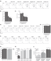
Comparison of mouse CD4+ T-cell isolation strategies. A–D) CD4+ T-cells were isolated from Foxp3-GFP mouse spleens using FACS sorting or different magnetic isolation strategies. Flow cytometry plots demonstrating the naïve CD4+ T-cell percentages (A), as well as bar graphs representing the total number (B), viability (C) and naïve CD4+ T-cell percentage (D) of isolated cells are shown. E–F) CD4+ T-cells were isolated from 5CC7-Foxp3 GFP-Rag2−/− mouse spleen. The flow cytometry plots (E) and bar graphs (F) representing the percentage of naïve CD4+ T-cells are shown. G–H) T-cells were isolated from Foxp3 GFP WT mouse spleen using either Naïve CD4+ T-cell Isolation Kit or different dilutions of depleting antibody cocktail. Flow cytometry plots (G) and bar graphs (H) demonstrating the naïve CD4+ T-cell purities are shown.
Fig. 2
Fig. 2
Comparison of iTreg yields and suppressive activities based on different initial CD4+ T-cell isolation strategies. A–C) T-cells isolated as described in Fig. 1A were stimulated for three days in order to generate iTregs. Flow cytometry plots (A) and bar graphs (B) representing the percentage of iTregs as well as bar graphs demonstrating the total number of iTregs (C) are shown. D–E) T-cells isolated as described in Fig. 1E were used to generate iTregs. iTreg purities are shown as flow cytometry plots (D) and as bar graphs (E). F–G) T cells isolated as described in Fig. 1G were cultured to generate iTregs. Flow cytometry plots (F) and bar graphs (G) represent the purity of iTregs. H–I) iTregs generated from T-cells obtained from WT FoxP3 GFP mice (H) and 5CC7-Foxp3 GFP-Rag2−/− mice (I) were tested using polyclonal and antigen specific suppression assays respectively. Percentage of suppression is shown as bar graphs.

References

    1. Akkaya B, Miozzo P, Holstein AH, Shevach EM, Pierce SK, Akkaya M. A simple, versatile antibody-based barcoding method for flow cytometry. J. Immunol. 2016;197:2027–2038. - PMC - PubMed
    1. Chattopadhyay G, Shevach EM. Antigen-specific induced T regulatory cells impair dendritic cell function via an IL-10/MARCH1-dependentmechanism. J. Immunol. 2013;191:5875–5884. - PMC - PubMed
    1. Chen W, Jin W, Hardegen N, Lei KJ, Li L, Marinos N, McGrady G, Wahl SM. Conversion of peripheral CD4+CD25− naive T cells to CD4+CD25+ regulatory T cells by TGF-beta induction of transcription factor Foxp3. J. Exp. Med. 2003;198:1875–1886. - PMC - PubMed
    1. Collison LW, Vignali DA. In vitro Treg suppression assays. Methods Mol. Biol. 2011;707:21–37. - PMC - PubMed
    1. Fantini MC, Dominitzki S, Rizzo A, Neurath MF, Becker C. In vitro generation of CD4+CD25+ regulatory cells from murine naive T cells. Nat. Protoc. 2007;2:1789–1794. - PubMed

Publication types

MeSH terms