. 2016 Dec 5;5(12):e004406.
doi: 10.1161/JAHA.116.004406.

Interleukin-37 and Dendritic Cells Treated With Interleukin-37 Plus Troponin I Ameliorate Cardiac Remodeling After Myocardial Infarction

Affiliations

Interleukin-37 and Dendritic Cells Treated With Interleukin-37 Plus Troponin I Ameliorate Cardiac Remodeling After Myocardial Infarction

Ruirui Zhu et al. J Am Heart Assoc. .

Abstract

Background: Excessive immune-mediated inflammatory reactions play a deleterious role in postinfarction ventricular remodeling. Interleukin-37 (IL-37) emerges as an inhibitor of both innate and adaptive immunity. However, the exact role of IL-37 and IL-37 plus troponin I (TnI)-treated dendritic cells (DCs) in ventricular remodeling after myocardial infarction (MI) remains elusive.

Methods and results: MI was induced by permanent ligation of the left anterior descending artery. Our results showed that treatment with recombinant human IL-37 significantly ameliorated ventricular remodeling after MI, as demonstrated by decreased infarct size, better cardiac function, lower mortality, restricted inflammatory responses, decreased myocardial fibrosis, and inhibited cardiomyocyte apoptosis. In vitro, we examined the phenotype of IL-37 plus TnI-conditioned DCs of male C57BL/6 mice and their capacity to influence the number of regulatory T cells. Our results revealed that IL-37 plus TnI-conditioned DCs obtained the characteristics of tolerogenic DCs (tDCs) and expanded the number of regulatory T cells when co-cultured with splenic CD4+ T cells. Interestingly, we also found that adoptive transfer of these antigen-loaded tDCs markedly increased the number of regulatory T cells in the spleen, attenuated the infiltration of inflammatory cells in the infarct hearts, decreased myocardial fibrosis, and improved cardiac function.

Conclusions: Our results reveal a beneficial role of IL-37 or tDCs treated with IL-37 plus TnI in post-MI remodeling that is possibly mediated by reestablishing a tolerogenic immune response, indicating that IL-37 or adoptive transfer of IL-37 plus TnI-treated tDCs may be a novel therapeutic strategy for ventricular remodeling after MI.

Keywords: Treg cells; interleukin‐37; myocardial infarction; remodeling; tolerogenic dendritic cells.


Figures

Figure 1
Figure 1
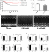
Interleukin (IL)–37 reduces myocardial infarct size and improves left ventricular function following myocardial infarction (MI). A, Survival analysis in PBS‐treated and IL‐37–treated mice after MI. B, Infarct size determined by 2, 3, 5‐triphenyltetrazolium chloride staining of sections on day 1 post‐MI (PBS‐treated group n=6 and IL‐37–treated group n=8). C, Representative M‐mode echocardiography images of the left ventricle on day 28 post‐MI. Left ventricular end‐diastolic dimension (LVEDD), left ventricular end‐systolic dimension (LVESD), ejection fraction (EF%), and fractional shortening (FS%) on day 7 (D) and day 28 (E) post‐MI (Sham n=6, PBS‐treated group n=6, and IL‐37‐treated group n=8). *P<0.05 and **P<0.01.
Figure 2
Figure 2
Changes in fibrosis and matrix metalloproteinase 2 (MMP2) expression after myocardial infarction (MI) in experimental groups. A, Representative Masson's trichrome staining images of collagen deposition (blue) in both infarct and remote areas on day 28 post‐MI. B, The extent of fibrosis, as assessed by the fibrotic area/left ventricle and interstitial fibrosis, was compared among the different groups (n=6 each). C, Representative images of Western blot demonstrating MMP2 expression in heart tissues on day 7 post‐MI (upper) and the data are presented as the fold change vs the sham group (lower) (n=6 each). Scale bar: 100 μm. **P<0.01.
Figure 3
Figure 3
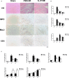
Inflammatory cells infiltration and cytokines expression in the infarcted heart. A, Representative images of haematoxylin and eosin (HE) staining, infiltration of myeloperoxidase (MPO+) neutrophils, mouse CD107b (Mac3+) macrophages, and CD3+ T cells in the border area of the infarct hearts. Images for neutrophils and macrophages are from day 3 after myocardial infarction (MI), and images for T cells are from day 7 post‐MI. B, Infiltration of neutrophils, macrophages, and T cells were compared between the different groups at set time points. C, Analysis of mRNA levels of proinflammatory cytokines interleukin (IL)–6, IL‐1β, and tumor necrosis factor‐α (TNF‐α), and anti‐inflammatory cytokines IL‐10 and transforming growth factor‐β (TGF‐β) on days 3 and 7 after MI. Data are depicted as fold changes vs sham. n=5 per group. Scale bar: 100 μm. **P<0.01 vs sham and # P<0.05, ## P<0.01 vs PBS+MI.
Figure 4
Figure 4
Interleukin (IL)–37 inhibited cardiomyocyte apoptosis in vivo. A, Representative images of terminal deoxynucleotidyl transferase dUTP nick‐end labeling (TUNEL)–stained heart sections from different groups 1 day and 28 days post–myocardial infarction (MI). TUNEL (green) and 4, 6‐diamidino‐2‐phenylindole (blue) staining of nuclei in apoptotic cardiomyocytes (red) in the peri‐infarct zone. B, Quantitative analysis of the percentages of TUNEL‐positive nuclei (n=6). Scale bar: 100 μm. **P<0.01.
Figure 5
Figure 5
Effects of interleukin (IL)–37 on regulatory T cells (Tregs), T helper (Th)1, and Th17 cells in spleens of C57BL/6 mice on day 7 after myocardial infarction (MI). A, CD4+ T‐cell subsets were gated, and representative images of Tregs, Th1, and Th17 cells are shown. B, Results of statistical analysis of average percentages of Tregs, Th1, and Th17 cells. C, Absolute numbers of Tregs, Th1, and Th17 cells were counted in the spleen. n=6 per group. APC indicates activated protein C; FITC, fluorescein isothiocyanate. **P<0.01 vs sham and ## P<0.01 vs PBS+MI.
Figure 6
Figure 6
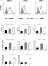
Interleukin (IL)–37 plus troponin I (TnI)–treated dendritic cells (DCs) display tolerogenic properties. A, Bone marrow–derived DCs (2×10⁵ cells/well) were cultured in the absence of stimulus (immature DCs [imDCs]) or in the presence of 1 μg/mL lipopolysaccharide (LPS) (mature DCs [mDCs]) or 10 ng/mL LPS, 30 ng/mL IL‐37, and 1 μg/mL TnIk (tolerogenic DCs [tDCs]). DCs were stained with isotype control antibodies or with specific antibodies against major histocompatibility complex class II (MHC‐II), CD40, and CD86 and analyzed by fluorescence‐activated cell sorting. B, Mean fluorescence intensities (MFIs) for MHCII, CD40, and CD86 were quantified. C, Analysis of the mRNA levels of IL‐10, transforming growth factor‐β (TGF‐β), indolamine 2, 3‐dioxygenase (IDO), interferon‐γ (IFN‐γ), and IL‐12 in different DCs groups. n=6 per group. **P<0.01.
Figure 7
Figure 7
Tolerogenic dentritic cells (tDCs) augment the percentage of regulatory T cells (Tregs) in vitro. A, Splenic CD4+ T cells (1×10⁶ cells/mL) were cultured for 3 days in the presence of medium alone or combined with immature DCs (imDCs), mature DCs (mDCs), or tDCs (2×10⁵ cells/mL). The cells were labeled 72 hours later with anti‐CD4, anti‐CD25, and anti‐Foxp3 and analyzed by fluorescence‐activated cell sorting (FACS). FACS data are representative of 1 of 6 to 8 independent experiments (each co‐culture preparation was prepared from a different mouse). B, Graphs represent the average percentages of Tregs (upper) and absolute numbers of Tregs (lower) in CD4+ splenic T cells of different groups. C, Analysis of the mRNA levels of Foxp3, interleukin (IL)–10, transforming growth factor‐β (TGF‐β), interferon‐γ (IFN‐γ), and IL‐17A in different groups. No DCs group n=6, imDCs group n=8, mDCs group n=8, and tDCs group n=8. **P<0.01.
Figure 8
Figure 8
Tolerogenic dentritic cells (tDCs) increase the number of regulatory T cells (Tregs) and decreases T helper (Th)1 and Th17 cells in spleen on day 7 after myocardial infarction (MI). A, CD4+ T‐cell subsets were gated, and representative images of Tregs, Th1, and Th17 cells are shown. B, Results of statistical analysis of average percentages of Tregs, Th1, and Th17 cells. C, Absolute numbers of Tregs, Th1, and Th17 cells were counted in the spleen. n=6 per group. **P<0.01.
Figure 9
Figure 9
Troponin I (TnI)–pulsed tolerogenic dentritic cells (tDCs) induced an antigen‐specific regulatory T (Treg) cell population. A, Mice received one intravenous injection of PBS, antigen‐unpulsed tolerogenic DCs (Un‐tDCs), TnI‐tDCs, or type II collagen tDCs (CII‐tDCs) (antigen‐mismatched tDCs) 24 hours after myocardial infarction (MI). Six days later, mice were sacrificed and splenocytes harvested. For Treg cell analysis by flow cytometry, the splenocytes were stained with anti‐CD4, anti‐CD25 and then anti‐Foxp3. The percentage and absolute numbers of CD4+ CD25+Foxp3+ cells are shown. B, Foxp3 mRNA levels from the splenocytes (cultured with TnI for 72 hours) were determined by real‐time polymerase chain reaction. C, Analysis of the mRNA levels of interferon‐γ (IFN‐γ) and interleukin (IL)–10 in different groups. n=6 per group. **P<0.01.
Figure 10
Figure 10
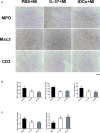
Tolerogenic dentritic cells (tDCs) decrease inflammatory cells infiltration in the infarcted heart. A, Representative images of infiltration of myeloperoxidase (MPO+) neutrophils, mouse CD107b (Mac3+) macrophages, and CD3+ T cells in the border area of the infarct hearts. Images for neutrophils and macrophages are from day 3 after myocardial infarction (MI), and images for T cells are from day 7 post‐MI. B, Infiltration of neutrophils, macrophages, and T cells were compared between the different groups. n=5 per group. Scale bar: 100 μm. **P<0.01.
Figure 11
Figure 11
Adoptive transfer of tolerogenic dendritic cells (tDCs) inhibited cardiac fibrosis and prevented left ventricular function following myocardial infarction (MI). A, Representative Masson's trichrome staining images of collagen deposition (blue) in the border area of the infarct hearts on day 28 post‐MI (upper), and representative M‐mode echocardiography images of the left ventricle (LV) on day 28 post‐MI (lower). B, The extent of fibrosis, as assessed by the fibrotic area/LV, was compared among the different groups. C, Data for left ventricular end‐diastolic dimension (LVEDD) and ejection fraction (EF%) in the 4 groups of mice. n=6 per group. **P<0.01.
Figure 12
Figure 12
Inflammatory cells infiltration and cytokines expression in the infarcted heart after treatment with interleukin (IL)‐37 or tolerogenic dendritic cells (tDCs) 24 hours post–myocardial infarction (MI). A, Representative images of infiltration of myeloperoxidase (MPO+) neutrophils, mouse CD107b (Mac3+) macrophages, and CD3+ T cells in the border area of the infarct hearts. Images for neutrophils and macrophages are from day 3 after MI, and images for T cells are from day 7 post‐MI. B, Infiltration of neutrophils, macrophages, and T cells were compared between the different groups. C, Analysis of mRNA levels of tumor necrosis factor‐α (TNF‐α) and IL‐10 on day 7 after MI. Data are depicted as fold changes vs PBS+MI. n=5 per group. Scale bar: 100 μm. **P<0.01 vs PBS+MI.

References

    1. Frantz S, Bauersachs J, Ertl G. Post‐infarct remodelling: contribution of wound healing and inflammation. Cardiovasc Res. 2009;81:474–481. - PMC - PubMed
    1. Nian M, Lee P, Khaper N, Liu P. Inflammatory cytokines and postmyocardial infarction remodeling. Circ Res. 2004;94:1543–1553. - PubMed
    1. Frangogiannis NG. The immune system and cardiac repair. Pharmacol Res. 2008;58:88–111. - PMC - PubMed
    1. Timmers L, Sluijter JP, van Keulen JK, Hoefer IE, Nederhoff MG, Goumans MJ, Doevendans PA, van Echteld CJ, Joles JA, Quax PH, Piek JJ, Pasterkamp G, de Kleijn DP. Toll‐like receptor 4 mediates maladaptive left ventricular remodeling and impairs cardiac function after myocardial infarction. Circ Res. 2008;102:257–264. - PubMed
    1. Zhou SF, Yuan J, Liao MY, Xia N, Tang TT, Li JJ, Jiao J, Dong WY, Nie SF, Zhu ZF, Zhang WC, Lv BJ, Xiao H, Wang Q, Tu X, Liao YH, Shi GP, Cheng X. IL‐17A promotes ventricular remodeling after myocardial infarction. J Mol Med (Berl). 2014;92:1105–1116. - PubMed

Publication types

MeSH terms