Review
. 2017 Mar 1;23(2):232-254.
doi: 10.1093/humupd/dmw042.

New concepts for an old problem: the diagnosis of endometrial hyperplasia

Affiliations
Review

New concepts for an old problem: the diagnosis of endometrial hyperplasia

Peter A Sanderson et al. Hum Reprod Update. .

Abstract

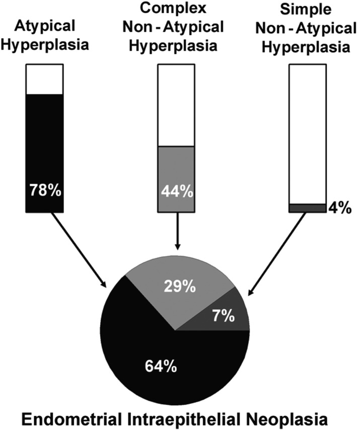
Background: Endometrial hyperplasia (EH) is a uterine pathology representing a spectrum of morphological endometrial alterations. It is predominantly characterized by an increase in the endometrial gland-to-stroma ratio when compared to normal proliferative endometrium. The clinical significance of EH lies in the associated risk of progression to endometrioid endometrial cancer (EC) and 'atypical' forms of EH are regarded as premalignant lesions. Traditional histopathological classification systems for EH exhibit wide and varying degrees of diagnostic reproducibility and, as a consequence, standardized patient management can be challenging.

Objective and rationale: EC is the most common gynaecological malignancy in developed countries. The incidence of EC is rising, with alarming increases described in the 40-44-year-old age group. This review appraises the current EH classification systems used to stratify women at risk of malignant progression to EC. In addition, we summarize the evidence base regarding the use of immunohistochemical biomarkers for EH and discuss an emerging role for genomic analysis.

Search methods: PubMed, Medline and the Cochrane Database were searched for original peer-reviewed primary and review articles, from January 2000 to January 2016. The following search terms were used: 'endometrial hyperplasia', 'endometrial intraepithelial neoplasia', 'atypical hyperplasia', 'complex atypical hyperplasia', 'biomarker', 'immunohistochemistry', 'progression', 'genomic', 'classification' and 'stratification'.

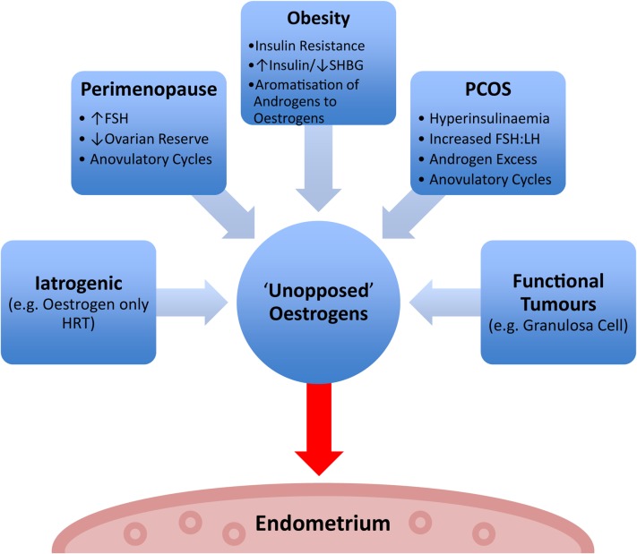
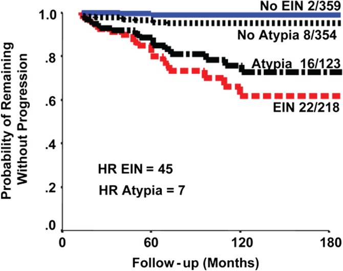
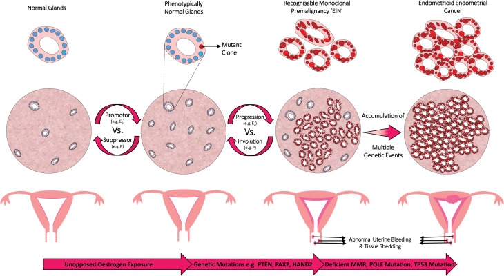
Outcomes: Recent changes to EH classification reflect our current understanding of the genesis of endometrioid ECs. The concept of endometrial intraepithelial neoplasia (EIN) as a mutationally activated, monoclonal pre-malignancy represents a fundamental shift from the previously held notion that unopposed oestrogenic stimulation causes ever-increasing hyperplastic proliferation, with accumulating cytological atypia that imperceptibly leads to the development of endometrioid EC. Our review highlights several key biomarker candidates that have been described as both diagnostic tools for EH and markers of progression to EC. We propose that, moving forwards, a 'panel' approach of combinations of the immunohistochemical biomarkers described in this review may be more informative since no single candidate can currently fill the entire role.

Wider implications: EC has historically been considered a predominantly postmenopausal disease. Owing in part to the current unprecedented rates of obesity, we are starting to see signs of a shift towards a rising incidence of EC amongst pre- and peri-menopausal woman. This creates unique challenges both diagnostically and therapeutically. Furthering our understanding of the premalignant stages of EC development will allow us to pursue earlier diagnosis and facilitate appropriate stratification of women at risk of developing EC, permitting timely and appropriate therapeutic interventions.

Keywords: biomarkers; endometrial hyperplasia; endometrial intraepithelial neoplasia; endometrioid endometrial cancer; genomic classification; immunohistochemistry; patient stratification; personalized medicine; progression.


Figures

Figure 1
Figure 1
Haematoxylin and eosin (H&E) stained sections demonstrating variation in size and shape of endometrial glands within a spectrum of endometrial hyperplasia (EH) lesions compared to proliferative endometrium (PE). Selection of glands marked by * in lumen. (A) PE, (B) hyperplasia without atypia: large cystically dilated glands, (C) endometrial intraepithelial neoplasia (EIN): varied and irregular gland morphology, (D) high-power EIN lesion: cytological atypia within glands (arrow) and (E) excerpt of a phenotypically ‘normal’ gland cytology within the same section as D for comparison. Varying magnifications: see scale bars.
Figure 2
Figure 2
Factors contributing to ‘unopposed’ oestrogen stimulation of the endometrium. SHBG = sex hormone binding globulin, FSH = follicle stimulating hormone, FSH:LH = follicle stimulating hormone to luteinizing hormone ratio, HRT = hormone replacement therapy, PCOS = polycystic ovarian syndrome.
Figure 3
Figure 3
Clonal expansion of EIN. H&E staining of an endometrial biopsy. (A) Low power view of a clonal expansion of EIN, with prominent gland crowding (marked in oval with bold dashes), in a background endometrium demonstrating benign EH, (B) high-power view of background endometrium, (C) high-power view of EIN lesion glands. Varying magnifications: see scale bars.
Figure 4
Figure 4
Correlation of WHO and EIN diagnoses (Hecht et al., 2005). (1) The bar graphs show the approximate percentage of each WHO94 category that would be considered as EIN. Residual WHO94 EHs that are not diagnostic of EIN (i.e. the white areas of the bars) are attributed to unopposed oestrogen (anovulatory cycles), polyps and other causes. (2) The pie chart demonstrates the relative contributions of each hyperplasia subtype to the EIN diagnostic category in a series of 97 cases with 28 EIN examples by Hecht et al. from their 2005 study. Republished with permission from Hecht et al. (2005).
Figure 5
Figure 5
Data to suggest that EIN classification system more accurately predicts progression to EC than the WHO94 system (Baak et al., 2005a). Patients with at least 1-year follow-up. The graph compares the WHO94 ‘Atypia’ and EIN systems in terms of prognostic accuracy. The study reported that the EIN classification is superior to the WHO94 classification for discerning cases at risk of progression to future EC. The fractions are the number that progressed to cancer over the total in that subgroup. HR = hazard ratio. Republished with permission from Baak et al. (2005a).
Figure 6
Figure 6
PTEN immunohistochemical staining of an EIN lesion. (A) H&E stained endometrial biopsy section demonstrating a region of EIN (below black line). (B) PTEN immunohistochemical staining of the same tissue section as in A. PTEN-null glands demonstrated by a loss of brown (DAB) cytoplasmic and nuclear staining in the same region corresponding to the EIN lesion as seen in image A. (Mouse monoclonal anti-human PTEN clone 6H2.1, Dako, Ely, UK; Antigen retrieval: decloaking chamber in citrate pH6; Overnight incubation 1:300 at 4°C.) Magnification: see scale bars.
Figure 7
Figure 7
PTEN immunohistochemical staining of hyperplasia without atypia. PTEN immunohistochemical staining demonstrating isolated PTEN-null glands (loss of brown (DAB) staining) seen within two separate tissue sections diagnosed as hyperplasia without atypia. (Mouse monoclonal anti-human PTEN clone 6H2.1, Dako, Ely, UK; antigen retrieval: decloaking chamber in citrate pH6; overnight incubation 1:300 at 4°C.) Magnification: see scale bars.
Figure 8
Figure 8
PAX2 immunohistochemical staining. Selection of glands marked by * in lumen. (A) PAX2 stained section of proliferative endometrium demonstrating strong brown nuclear (DAB) PAX2 staining within the glands, (B) loss of brown (DAB) PAX2 nuclear staining within the glands of an EIN lesion above/right of the black line. Crowded glandular background endometrium can also be seen. (Rabbit anti-PAX2 polyclonal Z-RX2, Invitrogen, Camarillo, CA; Antigen retrieval: decloaking chamber in citrate pH6; Overnight incubation 1:500 at 4°C.) Magnification: see scale bars.
Figure 9
Figure 9
Flow diagram detailing proposed initial management of EHs based upon guidance released by the Royal College of Obstetricians and Gynaecologists (RCOG), UK in 2016 (Gallos et al., 2016). *A comprehensive treatment review and plan for ongoing management should be made at this point dependant on the outcome of the preceding endometrial biopsy and in keeping with local and national guidelines/practise. aRisk factors for EH are specified in Table I. bOvarian conservation should be considered according to patients age, menopausal status and preferences. Total hysterectomy may also be indicated where there are (i) adverse effects with medical treatments, (ii) concerns over medication compliance and (iii) patient preference, e.g. elevated anxiety. cMedical progestin therapy has varying forms; for further recommendations, refer to national guidelines (Committee on Gynecologic Practice, 2015; Gallos et al., 2016). dFollow-up intervals for patients undergoing medical treatment of EIN should be tailored to individual patients and must reflect any ongoing risk factors, symptomatology and treatment responses. BSO = bilateral salpingo-oophorectomy.
Figure 10
Figure 10
A schematic diagram to illustrate a proposed mechanism for monoclonal development of EIN. The endometrium is exposed to unopposed oestrogens via several possible routes (as described in Fig. 2). Oestrogen (E2), acting as a promotor, drives proliferation of the endometrial glands. This process can be reversible, e.g. with progestin (P) therapy acting as a suppressor. In ‘at risk’ individuals, a mutant clone may develop in this environment. The mutant clone occurs within phenotypically normal appearing endometrial glands. The mutant clone is selected for and progresses, aided by the influence of unopposed oestrogens. Over time, with the accrual of further genetic damage, not yet fully elucidated (bottom arrow shows suggestions), the mutant clone proliferates and an EIN lesion can be diagnosed during routine light microscopic examination of an H&E stained section. Endocrine modifiers can alter the balance of EIN progression versus involution. The patient may present with symptoms of abnormal uterine bleeding (AUB) and a thickened endometrium on ultrasound imaging. With the continued accumulation of further genetic damage, not yet fully elucidated (bottom arrow shows suggestions), the EIN lesion undergoes malignant transformation to EC.

References

    1. Abu-Rustum NR, Zhou Q, Gomez JD, Alektiar KM, Hensley ML, Soslow RA, Levine DA, Chi DS, Barakat RR, Iasonos A. A nomogram for predicting overall survival of women with endometrial cancer following primary therapy: toward improving individualized cancer care. Gynecol Oncol 2010;116:399–403. - PMC - PubMed
    1. Ahmed ARH, Muhammad EMS. E-cadherin and CD10 expression in atypical hyperplastic and malignant endometrial lesions. J Egypt Natl Canc Inst 2014;26:211–217. - PubMed
    1. Allison KH, Tenpenny E, Reed SD, Swisher EM, Garica RL. Immunohistochemical markers in endometrial hyperplasia: is there a panel with promise. Appl Immunohistochem Mol Morphol 2008;16:329–343. - PubMed
    1. Allison KH, Upson K, Reed SD, Jordan CD, Newton KM, Doherty J, Swisher EM, Garcia RL. PAX2 loss by immunohistochemistry occurs early and often in endometrial hyperplasia. Int J Gynecol Pathol 2012;31:159–167. - PMC - PubMed
    1. Amant F, Moerman P, Neven P, Timmerman D, Van Limbergen E, Vergote I. Endometrial cancer. Lancet 2005;366:491–505. - PubMed