. 2017 Apr;28(4):1227-1238.
doi: 10.1681/ASN.2016050496. Epub 2016 Dec 5.

Toll-Like Receptor 9 Stimulation Induces Aberrant Expression of a Proliferation-Inducing Ligand by Tonsillar Germinal Center B Cells in IgA Nephropathy

Affiliations

Toll-Like Receptor 9 Stimulation Induces Aberrant Expression of a Proliferation-Inducing Ligand by Tonsillar Germinal Center B Cells in IgA Nephropathy

Masahiro Muto et al. J Am Soc Nephrol. 2017 Apr.

Abstract

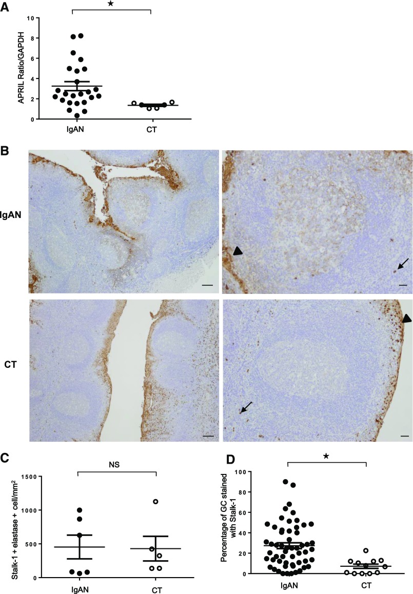
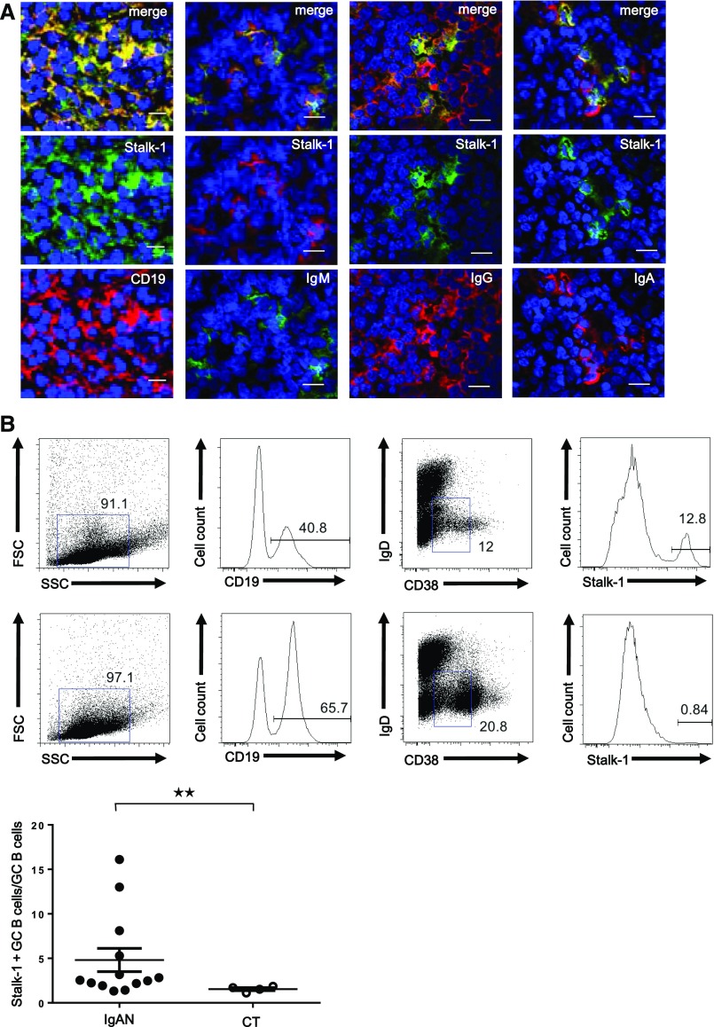
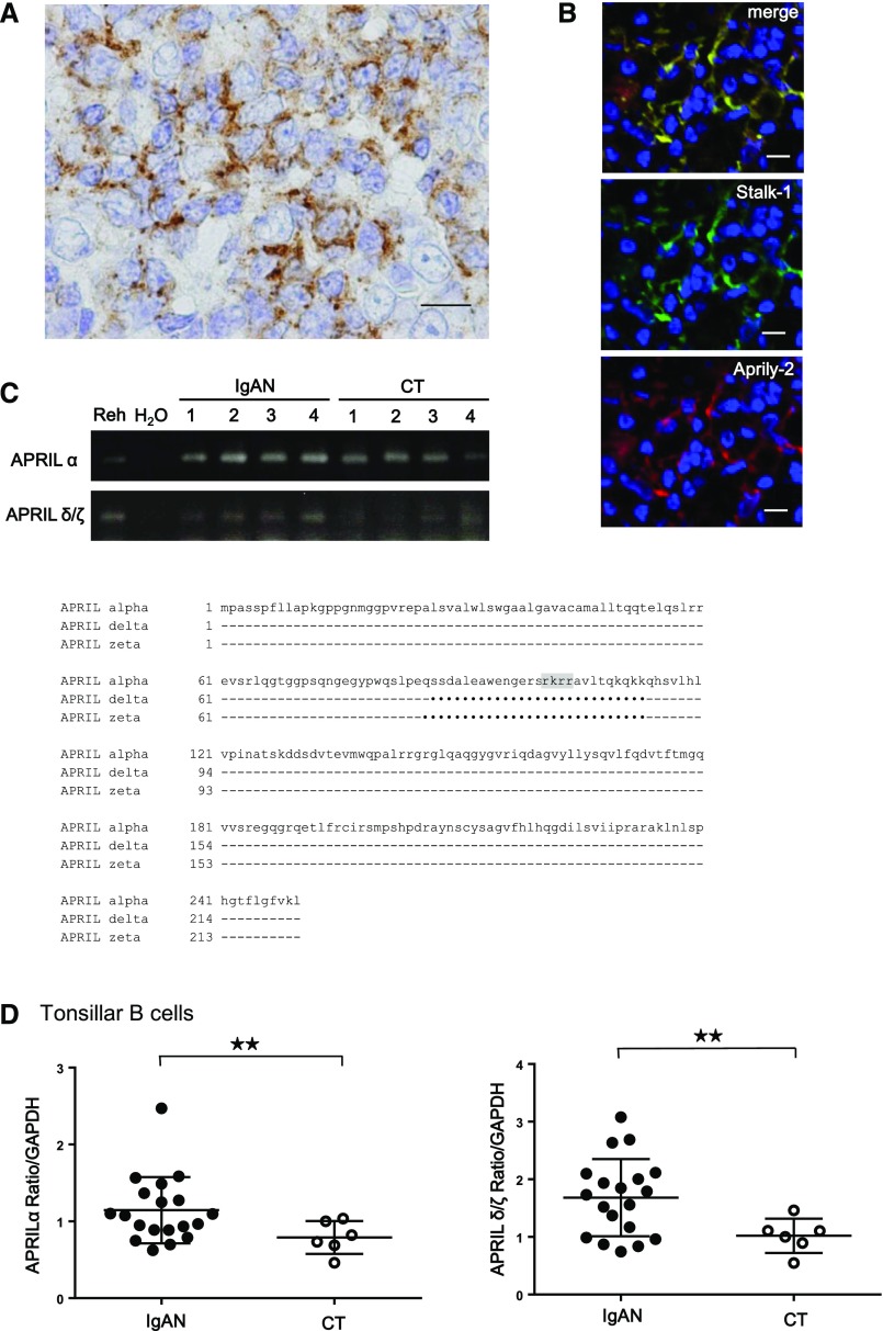
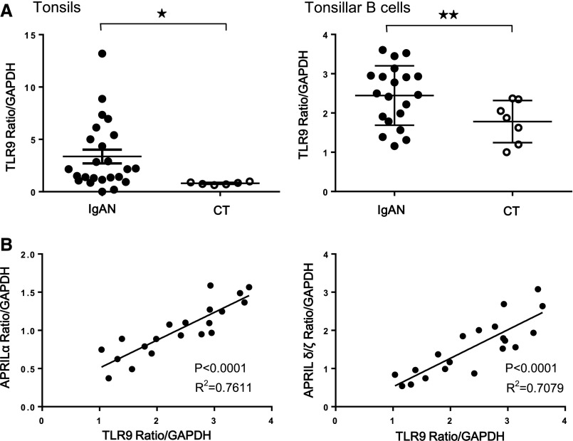
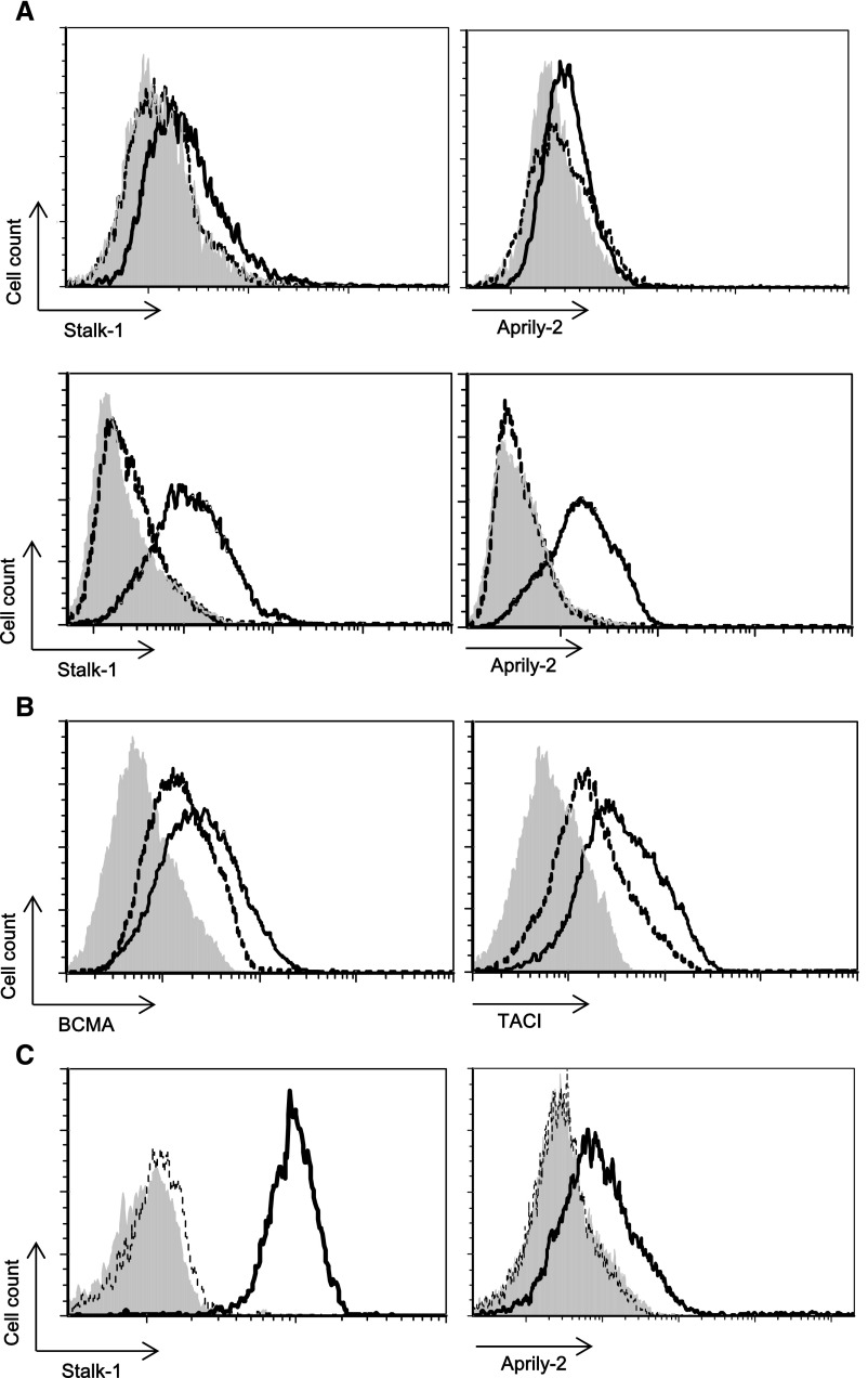
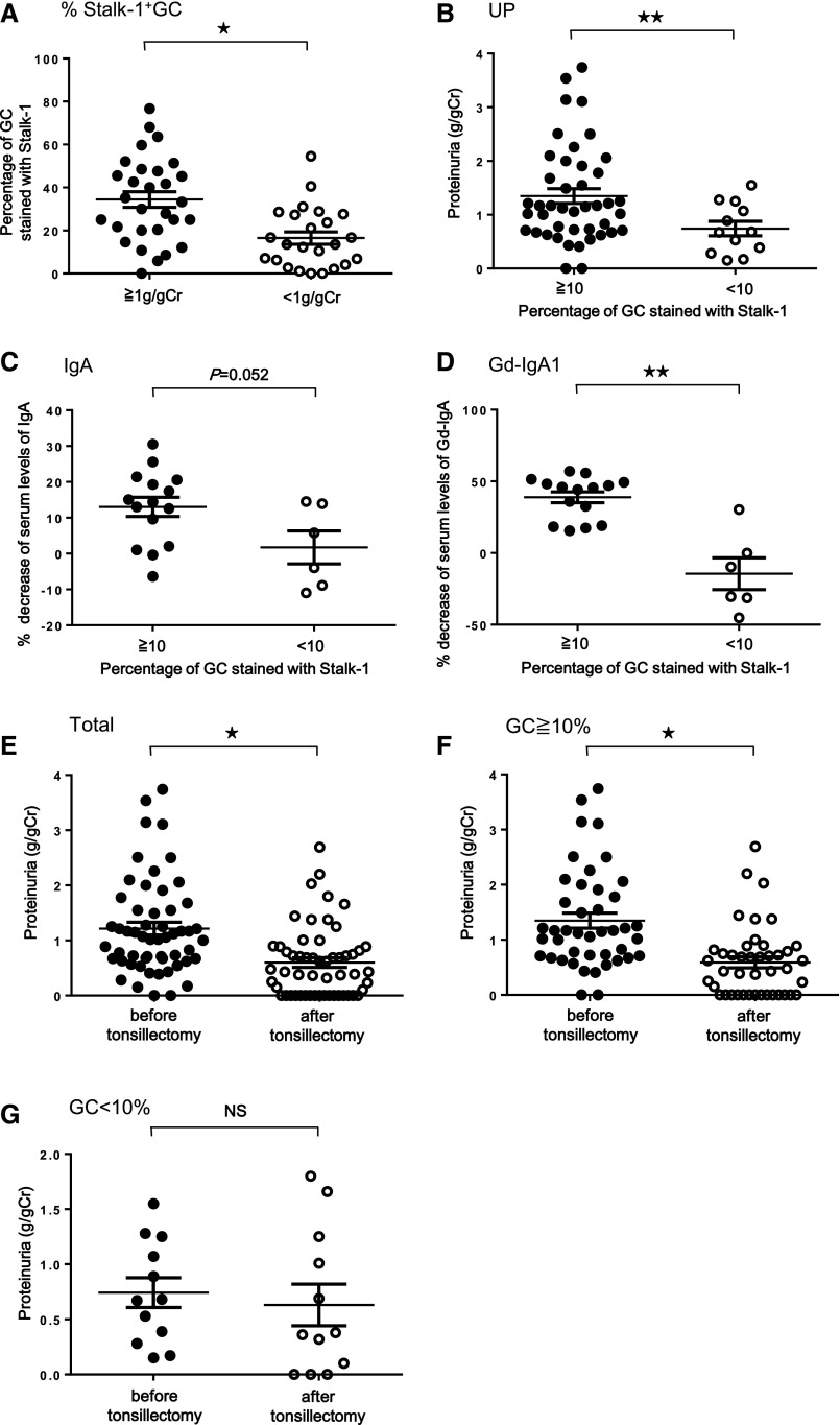
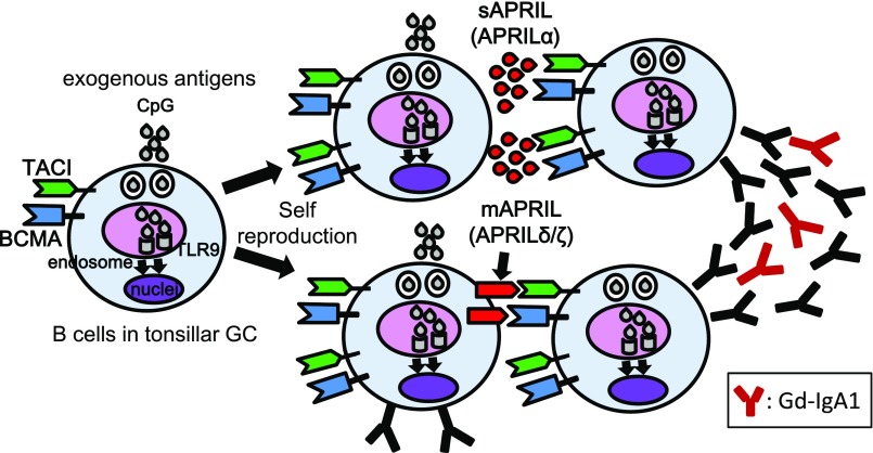
The TNF family member a proliferation-inducing ligand (APRIL; also known as TNFSF13), produced by myeloid cells, participates in the generation and survival of antibody-producing plasma cells. We studied the potential role of APRIL in the pathogenesis of IgA nephropathy (IgAN). We found that a significant proportion of germinal centers (GCs) in tonsils of patients with IgAN contained cells aberrantly producing APRIL, contributing to an overall upregulation of tonsillar APRIL expression compared with that in tonsils of control patients with tonsillitis. In IgAN GC, antigen-experienced IgD-CD38+/-CD19+ B cells expressing a switched IgG/IgA B cell receptor produced APRIL. Notably, these GC B cells expressed mRNA encoding the common cleavable APRIL-α but also, the less frequent APRIL-δ/ζ mRNA, which encodes a protein that lacks a furin cleavage site and is, thus, the uncleavable membrane-bound form. Significant correlation between TLR9 and APRIL expression levels existed in tonsils from patients with IgAN. In vitro, repeated TLR9 stimulation induced APRIL expression in tonsillar B cells from control patients with tonsillitis. Clinically, aberrant APRIL expression in tonsillar GC correlated with greater proteinuria, and patients with IgAN and aberrant APRIL overexpression in tonsillar GC responded well to tonsillectomy, with parallel decreases in serum levels of galactose-deficient IgA1. Taken together, our data indicate that antibody disorders in IgAN associate with TLR9-induced aberrant expression of APRIL in tonsillar GC B cells.

Keywords: IgA nephropathy; cytokines; immunology.


Figures

Figure 1.
Figure 1.
Increased APRIL expression in tonsils from patients with IgAN. (A) Tonsillar APRIL mRNA expressions in patients with IgAN (n=24) were significantly higher than those in patients with CT (n=6). Bars represent the mean±SEM. *P<0.01. (B) Immunohistochemistry with Stalk-1 (specific for APRIL-producing cells) in patients with IgAN (upper panels) and patients with CT (lower panels). Representative GCs are shown in right panels. Arrows and arrowheads mark Stalk-1–stained neutrophil and epithelial cells, respectively. Pictures shown are representative of 56 tonsils from patients with IgAN and 12 tonsils from patients with CT. Scale bars, 250 μm in left panels; 100 μm in right panels. (C) Quantification of Stalk-1+ elastase+ neutrophils showed no significant difference. (D) However, percentage of GC–containing APRIL–producing cells (Stalk-1+GC) was significantly different in total tonsillar GCs from patients with IgAN and patients with CT. *P<0.01.
Figure 2.
Figure 2.
CD19+ B cells produce APRIL in tonsillar GCs of patients with IgAN. (A) IgAN tonsils showed costaining for Stalk-1 (green) as well as CD19, IgM, IgG, and IgA (red). A representative GC is shown. Pictures shown are representative of tonsils from patients with IgAN. Scale bars, 20 μm. (B) A cell suspension from IgAN and CT tonsils was surface stained for CD19, CD38, IgD, and after cell permeabilization, Stalk-1 (top and middle panels). Plots for cells gated on CD19 are representative of 13 patients with IgAN and four patients with CT. The percentage of Stalk-1+ cells among CD19+IgDCD38+/− cells is also shown (bottom panel). **P<0.05.
Figure 3.
Figure 3.
Tonsillar GC B cells of IgAN express cleavable and uncleavable APRIL. (A) IgAN tonsils were stained for Stalk-1. A representative GC B cell is shown. The picture shown is representative of 56 patients with IgAN. (B) IgAN tonsils were costained for Stalk-1 (green) and Aprily-2 (red). A representative GC is shown. Scale bars, 20 μm. (C) Predicted amino acid sequences of different isoforms of APRIL. The GenBank accession numbers for APRIL-α, -δ, and -ζ are NM_003808, NM_001198622, and NM_001198623.1, respectively. The furin cleavable site lacking in APRIL-δ and -ζ is highlighted in gray. Identities are indicated by dashes, and deletions are indicated by dots. Numbers indicate amino acid positions. (D) Correlation between APRIL-α and -δ/ζ mRNA expression in purified tonsillar B cells from patients with IgAN (n=20) and patients with CT (n=6). Both APRIL-α and -δ/ζ mRNA expressions in tonsillar B cells were significantly higher in patients with IgAN. Bars represent the mean±SEM. **P<0.05.
Figure 4.
Figure 4.
Correlation between TLR9 and APRIL mRNA expressions in patients with IgAN. (A) TLR9 mRNA expressions in whole tonsils (left panel) and purified tonsillar B cells (right panel) were significantly higher in IgAN. Bars represent the mean±SEM. *P<0.01; **P<0.05. (B) TLR9 and APRIL-α (left panel) or -δ/ζ (right panel) mRNA expressions in tonsillar B cells were well correlated in patients with IgAN.
Figure 5.
Figure 5.
TLR9 activation induces APRIL expression in tonsillar B cells. (A) Tonsillar B cells isolated from patients with CT were stimulated daily with 10 μg/ml CpG. APRIL expression is shown on viable (upper panel) and permeabilized (lower panel) gated CD19+ B cells. (B) Surface expressions of TACI and BCMA are also shown. (C) CD19+ B cells from patients with CT were purified on an FACS ARIA (BD Pharmingen) by positive selection. Purified CD19+ B cells were stimulated daily with 10 μg/ml CpG. APRIL expression is shown on permeabilized cells. Shaded histograms represent control isotype–matched reactivity. Dotted and straight lines represent indicated antibody reactivities on control and CpG-ODN–stimulated cells, respectively, at day 7. Histogram plots are representative of at least three experiments performed with tonsils from independent patients.
Figure 6.
Figure 6.
Correlation between APRIL expression in tonsillar GC and disease activity in patients with IgAN. (A) Percentage of Stalk-1+GC in tonsils of patients with IgAN and their proteinuria level. The percentage of Stalk-1+GC in patients with IgAN with proteinuria >1 g/g creatinine (Cr) was significantly higher than that in those with proteinuria <1 g/g Cr. *P<0.01. (B) Comparison of proteinuria level, (C) percentage decrease of the serum IgA, and (D) the serum Gd-IgA1 levels between patients with IgAN with the percentage of Stalk-1+GC≥10% and those with the percentage of Stalk-1+GC<10%. Patients with IgAN with Stalk-1+GC≥10% showed significantly higher proteinuria before tonsillectomy and larger decrease of serum levels of Gd-IgA1 after tonsillectomy than those with Stalk-1+GC<10%. **P<0.05. (E–G) Comparison of proteinuria levels before and after tonsillectomy in (E) patients with IgAN (*P<0.01), (F) those with Stalk-1+GC≥10% (*P<0.01), and (G) those with Stalk-1+GC<10%. There were significant differences before and after tonsillectomy in the preceding two groups, despite no significance in patients with IgAN with Stalk-1+GC<10%. The average duration from tonsillectomy to quantification of these clinical parameters was 69.2±47.2 days.
Figure 7.
Figure 7.
Crosstalk between APRIL and TLR9 on B cells in tonsillar GCs of patients with IgAN. This study revealed aberrant APRIL expression in tonsillar GC B cells from patients with IgAN. On the basis of our findings, we hypothesize that activation of intracellular TLR9 through exogenous antigens may be involved in this overexpression consisting of not only APRIL-α but also, uncleaved APRIL, such as APRIL-δ/ζ in tonsillar GC B cells of patients with IgAN. This TLR9 activation also upregulates expression of TACI and BCMA and increases both BCR signaling and APRIL sensitivity. This aberrant APRIL expression may induce long-term survival of GC B cells responsible for the production of aberrant antibodies, including Gd-IgA1, and thereby, contribute to subsequent progression of IgAN.

Comment in

Similar articles

Cited by

References

    1. Barratt J, Feehally J: IgA nephropathy. J Am Soc Nephrol 16: 2088–2097, 2005 - PubMed
    1. Appel GB, Waldman M: The IgA nephropathy treatment dilemma. Kidney Int 69: 1939–1944, 2006 - PubMed
    1. Suzuki Y, Suzuki H, Sato D, Kajiyama T, Okazaki K, Hashimoto A, Kihara M, Yamaji K, Satake K, Nakata J, Aizawa M, Novak J, Tomino Y: Reevaluation of the mucosa-bone marrow axis in IgA nephropathy with animal models. Adv Otorhinolaryngol 72: 64–67, 2011 - PubMed
    1. Suzuki Y, Tomino Y: Potential immunopathogenic role of the mucosa-bone marrow axis in IgA nephropathy: Insights from animal models. Semin Nephrol 28: 66–77, 2008 - PubMed
    1. Kokubo T, Hiki Y, Iwase H, Horii A, Tanaka A, Nishikido J, Hotta K, Kobayashi Y: Evidence for involvement of IgA1 hinge glycopeptide in the IgA1-IgA1 interaction in IgA nephropathy. J Am Soc Nephrol 8: 915–919, 1997 - PubMed

Substances