. 2017 Feb 15;37(5):e00497-16.
doi: 10.1128/MCB.00497-16. Print 2017 Mar 1.

The MLKL Channel in Necroptosis Is an Octamer Formed by Tetramers in a Dyadic Process

Affiliations

The MLKL Channel in Necroptosis Is an Octamer Formed by Tetramers in a Dyadic Process

Deli Huang et al. Mol Cell Biol. .

Abstract

Oligomerization of the mixed-lineage kinase domain-like protein (MLKL) is essential for its cation channel function in necroptosis. Here we show that the MLKL channel is an octamer comprising two previously identified tetramers most likely in their side-by-side position. Intermolecule disulfide bonds are present in the tetramer but are not required for octamer assembly and necroptosis. MLKL forms oligomers in the necrosome and is then released from the necrosome before or during its membrane translocation. We identified two MLKL mutants that could not oligomerize into octamers, although they formed a tetramer, and also, one MLKL mutant could spontaneously form a disulfide bond-linked octamer. Subsequent analysis revealed that the tetramers fail to translocate to the plasma membrane and that the MLKL octamer formation depends on α-helices 4 and 5. While MLKL could be detected from outside the cells, its N- and C-terminal ends could not be detected, indicating that the MLKL octamer spans across the plasma membrane, leaving its N and C termini inside the cell. These data allowed us to propose a 180° symmetry model of the MLKL octamer and conclude that the fully assembled MLKL octamers, but not the previously described tetramers, act as effectors of necroptosis.

Keywords: MLKL; channel; necroptosis; octamer.


Figures

FIG 1
FIG 1
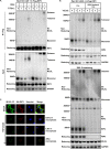
TNF-induced MLKL complex contains two disulfide bond-linked MLKL tetramers. (A) L929 cells were treated with TNF (10 ng/ml)-zVAD (20 μM) (T/Z) for 2 h, and MLKL fusion proteins were expressed in 293T cells. These cells were harvested, subjected to SDS-PAGE under nonreducing condition, and immunoblotted (IB) with anti-MLKL antibody. β-Me, β-mercaptoethanol; Ctrl, control. (B) L929 cells were treated with TNF-zVAD for the indicated times. These cells were lysed in the absence (−) or presence (+) of the irreversible chemical cross-linker DSS (0.5 mM) and incubated for 30 min at 4°C, and the cross-linking reactions were quenched by adding pH 7.5 Tris to a final concentration of 40 mM. Aliquots of the whole-cell lysates were subjected to SDS-PAGE under nonreducing conditions and immunoblotted with the indicated antibodies. *, nonspecific band. (C) L929 cells were treated or not treated with TNF-zVAD for 3 h, lysed in the absence or presence of DSS, and then fractionated. The TCL, crude membrane (Mem), and cytosol (Cyto) were immunoblotted under nonreducing conditions with antibodies against MLKL, RIP3, LDLR (a plasma membrane marker), and GAPDH. (D) Lysates of L929 cells treated or not treated with TNF-zVAD for 2 h were subjected to Superdex 200 gel filtration fractionation. The fractions were analyzed by immunoblotting under nonreducing conditions with antibody against MLKL. (E) Phosphomimic MLKL (with the S345D and S347D mutations, termed S2D) was expressed in Rip1 and Rip3 double-knockout L929 cells. The cells were lysed in the absence or presence of DSS. Aliquots of the whole-cell lysates were subjected to immunoblotting under nonreducing conditions with the indicated antibodies. L929 cell lysates were loaded as a control. (F) L929 cells were treated with TNF-zVAD in the presence or absence of GSK′872 (10 μM) for the indicated periods of time. These cells were lysed in the absence or presence of DSS, as described in the legend to panel B. Aliquots of the whole-cell lysates were subjected to immunoblotting under nonreducing conditions with the indicated antibodies. (G) WT RIP3 or the RIP3 T231A and S232A mutant (RIP3-2A) was expressed in Rip3 knockout L929 cells and stimulated with TNF-zVAD for 2 h. The cells were lysed in the absence or presence of DSS. Aliquots of the whole-cell lysates were subjected to immunoblotting under nonreducing conditions with the indicated antibodies. (H) Phosphorylated MLKL was generated by coexpression with the RIP3 kinase domain in Sf9 cells and then purified and subjected to gel filtration with Superdex 200. Fractions were collected, analyzed by SDS-PAGE, and stained by Coomassie blue.
FIG 2
FIG 2
The site of MLKL octamer formation is in the necrosome, and the octamers are released from the necrosome to function. (A) Rip3 KO L929 cells reconstituted with 3× Flag-RIP3 expression were treated with TNF-zVAD for different periods of time, as indicated. The cells were lysed in the absence (−) or presence (+) of DSS. RIP3 was immunoprecipitated (IP) with anti-Flag M2 beads. The cell lysates (Input) and immune complexes were analyzed by immunoblotting under nonreducing and reducing conditions. (B) Cells were treated and lysed as described in the legend to panel A. The RIP3 in half of the lysates was immunodepleted by immunoprecipitation with anti-Flag antibody M2 beads. TCL- and RIP3-depleted TCL were analyzed by immunoblotting under nonreducing condition using the indicated antibodies. *, nonspecific band. (C) Rip3 and Mlkl double-knockout L929 cells reconstituted with the expression of HA-RIP3 and MLKL-3×Flag were treated or not treated with TNF-zVAD for different times, as indicated, and then immunostained with anti-Flag antibody and anti-HA antibody and counterstained with Hoechst stain.
FIG 3
FIG 3
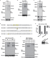
C169 and C275 form disulfide bonds in the MLKL tetramer, and the disulfide bond is not required for MLKL complex formation and is dispensable for TNF-induced necroptosis. (A) C-terminal 3×Flag-tagged WT MLKL or the indicated MLKL cysteine mutant was expressed in Mlkl KO L929 cells. The cells were treated or not treated with TNF-zVAD for 2 h and lysed. The cell lysates were analyzed by immunoblotting under nonreducing conditions with anti-MLKL antibody. (B) The cells described in the legend to panel A were treated or not treated with TNF-zVAD for 3 h. Viabilities were measured by determination of PI exclusion. The data represent the means ± SDs from triplicate experiments. (C) C-terminal 3×Flag-tagged WT, C169S MLKL, or C275S MLKL was expressed in Mlkl KO L929 cells. The cells were treated or not treated with TNF-zVAD for 2 h, lysed in the absence (−) or presence (+) of DSS, and immunoblotted with anti-MLKL and anti-RIP3 antibodies under nonreducing conditions. *, nonspecific band; DMSO, dimethyl sulfoxide. (D) The cells described in the legend to panel C were treated with TNF-zVAD for 2 h. RIP3 was immunoprecipitated with anti-Flag M2 beads. The cell lysates (Input) and immune complexes were analyzed by immunoblotting with the indicated antibodies. (E) The cells described in panel C were treated or not treated with TNF-zVAD for 2 h and then immunostained with anti-Flag antibody and PI and counterstained with Hoechst stain.
FIG 4
FIG 4
Human MLKL C86 is responsible for the formation of the disulfide bond between two human MLKL tetramers in vitro. (A) HeLa cells expressing human HA-RIP3 were treated or not treated with TNF-Smac-zVAD (T/S/Z; 30 ng/ml, 100 nM, and 20 μM, respectively) for 3 h. The cells were lysed in the absence (−) or presence (+) of DSS. Aliquots of the whole-cell lysates were subjected to immunoblotting under nonreducing condition with the indicated antibodies. (B) HeLa cells expressing human HA-RIP3 were treated or not treated human with TNF-Smac-zVAD for 3 h. Then, cell lysates were kept at 4°C overnight (lane II) or directly added with sample buffer (lanes I) before immunoblotting with anti-human MLKL antibody under nonreducing conditions. (C) L929 cells were treated with TNF-zVAD for 3 h. Then, cell lysates were kept at 4°C overnight (lane II) or directly added with sample buffer (lanes I) before immunoblotting with MLKL antibody under nonreducing conditions. (D) Alignment of the human MLKL N-terminal sequence and the mouse MLKL N-terminal sequence. (E) MLKL KO HeLa cells stably expressing RIP3 were reconstituted with the expression of human MLKL or MLKL C86S mutants. Cells were treated with human TNF-Smac-zVAD for 3 h. Then, the cell lysates were kept at 4°C overnight (lanes II) or directly added with sample buffer (lanes I) before immunoblotting with anti-human MLKL antibody under nonreducing conditions. (F) MLKL KO HeLa cells stably expressing RIP3 were reconstituted with the expression of human MLKL C86S mutants. Cells were treated or not treated with TNF-Smac-zVAD for 3 h. The cells were lysed in the absence or presence of DSS. Aliquots of the whole-cell lysates were subjected to immunoblotting under nonreducing conditions with the indicated antibodies. (G) The cells described in the legend to panel E were treated with TNF-zVAD for 3 h, and their viability was analyzed by determination of PI exclusion. The data represent the means ± SDs from triplicate experiments. The expression of MLKL and the MLKL C86S mutant was measured by immunoblotting with anti-MLKL antibody.
FIG 5
FIG 5
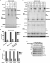
MLKL N-terminal α-helix 4 and α-helix 5 participate in the interaction between the two tetramers in the octamer. (A) Mlkl KO L929 cells reconstituted with the expression of C-terminal 3×Flag-tagged WT, S79A and K81A, and R105A and D106A MLKL. The cells were treated or not treated with TNF-zVAD for 2 h, lysed in the absence (−) or presence (+) of DSS, and immunoblotted with anti-MLKL and anti-RIP3 antibodies under nonreducing conditions. *, nonspecific band. (B) WT MLKL or the indicated MLKL mutant was expressed in Mlkl KO L929 cells. The cells were treated or not treated with TNF-zVAD for 2 h, lysed in the absence or presence of DSS, and immunoblotted with anti-MLKL antibody under nonreducing condition. (C) The cells described in the legend to panel A were treated or not treated with TNF-zVAD for 2 h, immunostained with anti-Flag antibody and PI, and counterstained with Hoechst stain. (D, E) The cells described in the legend to panel A or B were treated or not treated with TNF-zVAD for 3 h, and viabilities were measured by determination of PI exclusion. The data represent the means ± SDs from triplicate experiments. (F) The cells described in the legend to panel A were treated with TNF-zVAD for 2 h. RIP3 was immunoprecipitated with anti-Flag M2 beads. The cell lysates (Input) and immune complexes were analyzed by immunoblotting with the indicated antibodies.
FIG 6
FIG 6
MLKL N-terminal α-helix 5 is involved in the formation of the octamer. (A) Mlkl KO L929 cells reconstituted with the expression of WT MLKL, T122C MLKL, or T122C, C18S, C24S, and C28S MLKL were treated with TNF-zVAD for 2 h and then analyzed by immunoblotting with anti-MLKL antibody under nonreducing conditions. (B) The cells described in the legend to panel A were treated or not treated with TNF-zVAD for 3 h, and viabilities were measured by determination of PI exclusion. The data represent the means ± SDs from triplicate experiments. (C) Mlkl KO L929 cells reconstituted with the expression of WT, T122C, T122S, or T122A MLKL. The cells were treated with TNF-zVAD for 2 h, lysed in the absence (−) or presence (+) of DSS, and then analyzed by immunoblotting with anti-MLKL and anti-RIP3 antibodies under nonreducing conditions. *, nonspecific band. (D) The cells described in the legend to panel C were treated with TNF-zVAD for 3 h, and viabilities were measured by determination of PI exclusion. The data represent the means ± SDs from triplicate experiments. (E) The cells described in the legend to panel C were treated with TNF-zVAD for 2 h. RIP3 was immunoprecipitated with anti-Flag M2 beads. The cell lysates (Input) and immune complexes were analyzed by immunoblotting with the indicated antibodies.
FIG 7
FIG 7
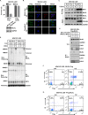
MLKL in the plasma membrane is a transmembrane protein, and both the N- and C-terminal ends are on the inner side of the membrane. (A) (Top) Mlkl KO L929 cells reconstituted with the expression of N-terminus-tagged MLKL (Flag-MLKL) or C-terminus-tagged MLKL (MLKL-Flag) were treated or not treated with TNF-zVAD for 3 h, and the viability of these cells was measured by determination of PI exclusion. The data represent the means ± SDs from triplicate experiments. (Bottom) The expression level of MLKL was measured by Western blotting. (B) The cells described in the legend to panel A were treated or not treated with TNF-zVAD for 2 h, immunostained with anti-Flag antibody and PI, and counterstained with Hoechst stain. (C) The cells described in the legend to panel A were treated with TNF-zVAD for 2 h. RIP3 was immunoprecipitated with anti-Flag M2 beads. The cell lysates (Input) and immune complexes were analyzed by immunoblotting with the indicated antibodies. P-MLKL, phosphorylated MLKL. (D) The cells described in the legend to panel A were treated or not treated with TNF-zVAD for 2 h, lysed in the absence (−) or presence (+) of DSS, and immunoblotted under nonreducing conditions with anti-MLKL and anti-RIP3 antibodies. *, nonspecific band. (E) Mlkl KO L929 cells expressing Flag-MLKL were treated or not treated with TNF-zVAD for 2 h and then labeled or not labeled with the membrane-impermeant reagent EZ-Link sulfo-NHS-LC-biotin for 20 min at 4°C. Then, these cells were lysed and the EZ-Link sulfo-NHS-LC-biotin-labeled proteins were pulled down by avidin beads at 4°C overnight. The cell lysates and pulldown complexes were immunoblotted with antibodies against MLKL, GAPDH, CAV1 (an inner membrane marker), and LDLR (an outer membrane marker). (F, G) Mlkl KO L929 cells expressing MLKL-Flag (F) or Flag-MLKL (G) were treated or not treated with TNF-zVAD for 2 h and then subjected to flow cytometry analysis of Flag and PI.
FIG 8
FIG 8
Model of MLKL octamer formation.

References

    1. Holler N, Zaru R, Micheau O, Thome M, Attinger A, Valitutti S, Bodmer JL, Schneider P, Seed B, Tschopp J. 2000. Fas triggers an alternative, caspase-8-independent cell death pathway using the kinase RIP as effector molecule. Nat Immunol 1:489–495. doi:10.1038/82732. - DOI - PubMed
    1. Zhang DW, Shao J, Lin J, Zhang N, Lu BJ, Lin SC, Dong MQ, Han J. 2009. RIP3, an energy metabolism regulator that switches TNF-induced cell death from apoptosis to necrosis. Science 325:332–336. doi:10.1126/science.1172308. - DOI - PubMed
    1. He S, Wang L, Miao L, Wang T, Du F, Zhao L, Wang X. 2009. Receptor interacting protein kinase-3 determines cellular necrotic response to TNF-α. Cell 137:1100–1111. doi:10.1016/j.cell.2009.05.021. - DOI - PubMed
    1. Cho Y, Challa S, Moquin D, Genga R, Ray TD, Guildford M, Chan FK-M. 2009. Phosphorylation-driven assembly of the RIP1-RIP3 complex regulates programmed necrosis and virus-induced inflammation. Cell 137:1112–1123. doi:10.1016/j.cell.2009.05.037. - DOI - PMC - PubMed
    1. Degterev A, Hitomi J, Germscheid M, Ch'en IL, Korkina O, Teng X, Abbott D, Cuny GD, Yuan C, Wagner G, Hedrick SM, Gerber SA, Lugovskoy A, Yuan J. 2008. Identification of RIP1 kinase as a specific cellular target of necrostatins. Nat Chem Biol 4:313–321. doi:10.1038/nchembio.83. - DOI - PMC - PubMed

Publication types

LinkOut - more resources