Endoscopic Removal of a Duodenal-Perforating Leg of Glasses with Dormia Basket
- PMID: 27920661
- PMCID: PMC5126602
- DOI: 10.1159/000452205
Endoscopic Removal of a Duodenal-Perforating Leg of Glasses with Dormia Basket
Abstract
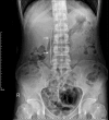
Ingestion of foreign bodies is common in clinical practice. Most ingested foreign bodies will pass through the gastrointestinal (GI) tract without any problems. While GI tract injury due to the ingested foreign body such as a toothpick, a fishbone, a date pit, or a chicken bone, is common, duodenal perforation is rare. In this report, our experience with this rare entity is shared. We present a 38-year-old male patient with GI tract perforation in the bulbus of the duodenum due to a leg of glasses. The patient was admitted to our hospital with severe abdominal pain. Right upper quadrant tenderness was detected at physical examination, and leukocytosis on the laboratory test results. Plain X-ray and computerized tomography showed an ingested foreign body in the bulbus of the duodenum. A leg of glasses perforating the duodenum was removed with endoscopy. The patient was managed nonoperatively, and discharged without any complications on the eighth day after endoscopy. Endoscopic removal and nonoperative management may be feasible in carefully selected patients with duodenal-perforating foreign bodies.
Keywords: Duodenal perforation; Endoscopic removal; Foreign body.
Figures


References
-
- Webb WA. Management of foreign bodies of the upper gastrointestinal tract. Gastroenterology. 1988;94:204–216. - PubMed
-
- Chaves DM, Ishioka S, Félix VN, et al. Removal of a foreign body from the upper gastrointestinal tract with a flexible endoscope: a prospective study. Endoscopy. 2004;36:887–892. - PubMed
-
- Birk M, Bauerfeind P, Deprez PH, et al. Removal of foreign bodies in the upper gastrointestinal tract in adults: European Society of Gastrointestinal Endoscopy (ESGE) Clinical Guideline. Endoscopy. 2016;48:489–496. - PubMed
-
- Goh BK, Chow PK, Quah HM, Ong HS, Eu KW, Ooi LL, et al. Perforation of the gastrointestinal tract secondary to ingestion of foreign bodies. World J Surg. 2006;30:372–377. - PubMed
Publication types
LinkOut - more resources
Full Text Sources
Other Literature Sources

