. 2016 Nov 22:7:1872.
doi: 10.3389/fmicb.2016.01872. eCollection 2016.

Transcriptional Profiling of Type II Toxin-Antitoxin Genes of Helicobacter pylori under Different Environmental Conditions: Identification of HP0967-HP0968 System

Affiliations

Transcriptional Profiling of Type II Toxin-Antitoxin Genes of Helicobacter pylori under Different Environmental Conditions: Identification of HP0967-HP0968 System

María G Cárdenas-Mondragón et al. Front Microbiol. .

Abstract

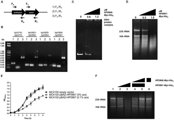
Helicobacter pylori is a Gram-negative bacterium that colonizes the human gastric mucosa and is responsible for causing peptic ulcers and gastric carcinoma. The expression of virulence factors allows the persistence of H. pylori in the stomach, which results in a chronic, sometimes uncontrolled inflammatory response. Type II toxin-antitoxin (TA) systems have emerged as important virulence factors in many pathogenic bacteria. Three type II TA systems have previously been identified in the genome of H. pylori 26695: HP0315-HP0316, HP0892-HP0893, and HP0894-HP0895. Here we characterized a heretofore undescribed type II TA system in H. pylori, HP0967-HP0968, which is encoded by the bicistronic operon hp0968-hp0967 and belongs to the Vap family. The predicted HP0967 protein is a toxin with ribonuclease activity whereas HP0968 is an antitoxin that binds to its own regulatory region. We found that all type II TA systems were expressed in H. pylori during early stationary growth phase, and differentially expressed in the presence of urea, nickel, and iron, although, the hp0968-hp0967 pair was the most affected under these environmental conditions. Transcription of hp0968-hp0967 was strongly induced in a mature H. pylori biofilm and when the bacteria interacted with AGS epithelial cells. Kanamycin and chloramphenicol considerably boosted transcription levels of all the four type II TA systems. The hp0968-hp0967 TA system was the most frequent among 317 H. pylori strains isolated from all over the world. This study is the first report on the transcription of type II TA genes in H. pylori under different environmental conditions. Our data show that the HP0967 and HP0968 proteins constitute a bona fide type II TA system in H. pylori, whose expression is regulated by environmental cues, which are relevant in the context of infection of the human gastric mucosa.

Keywords: H. pylori; HP0967; HP0968; environmental cues; toxin–antitoxin system.


Figures

FIGURE 1
FIGURE 1
HP0967–HP0968 is a bona fide type II TA system. (A) Schematic representation of the type II TA operons amplified for RT-PCR analysis using specific primers for toxin and antitoxin genes (arrows). (B) Qualitative RT-PCR assays carried out with RNA extracted from Helicobacter pylori 26695 grown in Brucella broth during 48 h. This is a representative result of two independent experiments. (C) EMSA exemplifying the binding of HP0968 Myc-His6 to hp0968–hp0967 promoter region. DNA-protein complex is indicated in the figure. (D) Ribonuclease assay using amounts of HP0967-Myc-His6 toxin with 1 μg total RNA. 23S rRNA and 16S rRNA are shown in the figure. (E) Growth kinetic of wild-type Escherichia coli (MC4100) harboring the empty vector pBAD-Myc-HisA and the pBAD-HP0967-Myc-His6 plasmid without and with induction of L-arabinose (ara). Bacterial cultures were grown for 8 h in LB medium at 37°C. (F) Ribonuclease inhibition assay using HP0967-Myc-His6 toxin in presence of HP0968-Myc-His6 antitoxin with 1 μg of total RNA. Alone RNA (lane 1); RNA with 0.5 (lane 2) and 1.0 μM (lane 3) of HP0967-Myc-His6 toxin; RNA with 1.0 μM of HP0967-Myc-His6 toxin and in presence of 0.6 (lane 4) and 1.2 μM (lane 5) of HP0968-Myc-His6 antitoxin; RNA with 1.2 μM of HP0968-Myc-His6 antitoxin (lane 6).
FIGURE 2
FIGURE 2
Environmental cues and expression of type II TA genes. Expression (qRT-PCR) of the type II TA genes (A) and cagA/vacA (B) of H. pylori 26695 in exponential (black bars) and stationary phase (gray bars). Fold-change expression (qRT-PCR) of the type II TA genes (C–F) and cagA/vacA (G) under different environmental conditions with respect to Brucella broth at 48 h. Data represent the mean of at least three independent experiments (mean ± SD). ns, not significant; statistically significant ∗∗∗p < 0.001; ∗∗p < 0.01; p < 0.05.
FIGURE 3
FIGURE 3
Expression of type II TA genes in biofilm formation and in AGS cells. (A) Quantification of biofilm formation of H. pylori 26695 by measuring Crystal Violet uptake at different times. Fold-change expression (qRT-PCR) of the type II TA genes and cagA/vacA in a mature biofilm after 72 h (B) and in contact with AGS cells (C). Data represent the mean of at least three independent experiments (mean ± SD). ns, not significant; statistically significant ∗∗∗p < 0.001; ∗∗p < 0.01; p < 0.05.
FIGURE 4
FIGURE 4
Effect of antibiotics on type II TA expression. Fold-change expression (qRT-PCR) of the type II TA genes (A–D) and cagA/vacA (E) in presence of different antibiotics concentrations such as: Ap (Ampicillin, 100 μg/ml), Km (Kanamycin, 30 μg/ml), Cm (Chloramphenicol, 30 μg/ml), and Tc (Tetracycline, 10 μg/ml) by 1 h. Data represent the mean of at least three independent experiments (mean ± SD). ns, not significant; statistically significant ∗∗∗p < 0.001; ∗∗p < 0.01; p < 0.05.
FIGURE 5
FIGURE 5
Prevalence of type II TA genes. (A) The clinical isolates tested (n = 114) comprised strains of H. pylori with different pathologies. Bars show the prevalence of type II TA genes in clinic (black) and sequenced (gray) strains. The heatmaps and hierarchical clustering of selected genomes for H. pylori and Helicobacter species showing both the presence (B) and identity (C) of toxin–antitoxin genes were determined in R (version 3.2.4) using the hclust function with the “ward.D” method. The red and blue colors indicate the presence of the toxin and the anti-toxin gene.

References

    1. Allan E., Clayton C. L., Mclaren A., Wallace D. M., Wren B. W. (2001). Characterization of the low-pH responses of Helicobacter pylori using genomic DNA arrays. Microbiology 147 2285–2292.10.1099/00221287-147-8-2285 - DOI - PubMed
    1. Altschul S. F., Madden T. L., Schaffer A. A., Zhang J., Zhang Z., Miller W., et al. (1997). Gapped BLAST and PSI-BLAST: a new generation of protein database search programs. Nucleic Acids Res. 25 3389–3402. 10.1093/nar/25.17.3389 - DOI - PMC - PubMed
    1. Amieva M. R., El-Omar E. M. (2008). Host-bacterial interactions in Helicobacter pylori infection. Gastroenterology 134 306–323. 10.1053/j.gastro.2007.11.009 - DOI - PubMed
    1. Aranda P. S., Lajoie D. M., Jorcyk C. L. (2012). Bleach gel: a simple agarose gel for analyzing RNA quality. Electrophoresis 33 366–369. 10.1002/elps.201100335 - DOI - PMC - PubMed
    1. Arcus V. L., Mckenzie J. L., Robson J., Cook G. M. (2011). The PIN-domain ribonucleases and the prokaryotic VapBC toxin-antitoxin array. Protein Eng. Des. Sel. 24 33–40. 10.1093/protein/gzq081 - DOI - PubMed

LinkOut - more resources