

Zinner syndrome-a rare developmental anomaly of the mesonephric duct diagnosed on magnetic resonance imaging
- PMID: 27920851
- PMCID: PMC5128193
- DOI: 10.1016/j.radcr.2016.04.002
Zinner syndrome-a rare developmental anomaly of the mesonephric duct diagnosed on magnetic resonance imaging
Abstract
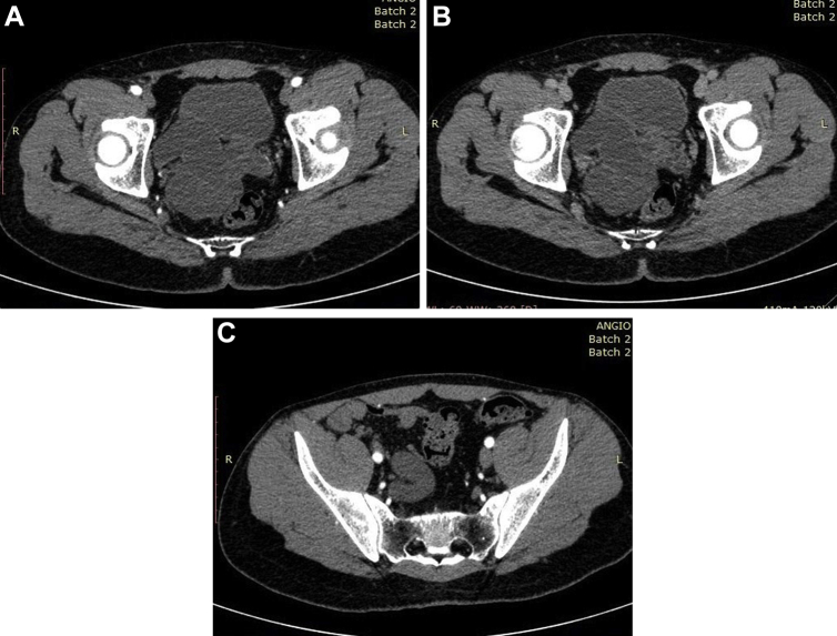
Developmental anomalies of the urogenital tract are rare but often encountered. Zinner's syndrome is a rare congenital abnormality of mesonephric (Wolffian) duct consisting of unilateral renal agenesis, ipsilateral seminal vesicle cyst, and ipsilateral ejaculatory duct obstruction due to developmental arrest in early embryogenesis affecting the caudal end of Mullerian duct and only approximately a 100 cases have been reported so far. Radiologic modalities such as intravenous pyelography, ultrasonography, vasovesiculography, contrast enhanced computed tomography, and magnetic resonance imaging are all helpful in diagnosis of this unusual entity. We present here an extremely rare developmental anomaly involving the Mullerian ducts, which would remain undiagnosed but for radiologic imaging. The patient presented with symptoms of lower urinary tract irritation.
Keywords: CT; MRI; USG; atretic ureter; ejaculatory duct; seminal vesicles.
Figures


References
-
- Pereira B.J., Sousa L., Azhinais P., Conceicao P., Borges R., Leao R. Zinner's syndrome: an up-to-date review of literature based on clinical case. Andrologia. 2009;41(5):322–330. - PubMed
-
- Zinner A. Ein fall von intravesikaler Samenblasenzyste. Wien Med Wochenschr. 1914;64:605.
-
- King B.F., Hattery R.R., Lieber M.M., Berquist T.H., Williamson B., Jr., Hartman G.W. Congenital cystic disease of the seminal vesicle. Radiology. 1991;178:207–211. - PubMed
-
- Gianna P., Giuseppe P.G. Mayer-Rokitansky-Küster-Hauser syndrome and the Zinner syndrome, female and male malformation of reproductive system: are two separate entities? J Chinese Clin Med. 2007;2:11.
-
- Livingston L., Larson C.R. Seminal vesicle cyst with ipsilateral renal agenesis. AJR Am J Roentgenol. 2000;175:177–180. - PubMed
Publication types
LinkOut - more resources
Full Text Sources
Other Literature Sources
