Review
. 2016 Dec 6;6(6):20160055.
doi: 10.1098/rsfs.2016.0055.

Recent advances in the preparation and application of multifunctional iron oxide and liposome-based nanosystems for multimodal diagnosis and therapy

Affiliations
Review

Recent advances in the preparation and application of multifunctional iron oxide and liposome-based nanosystems for multimodal diagnosis and therapy

Marzia Marciello et al. Interface Focus. .

Abstract

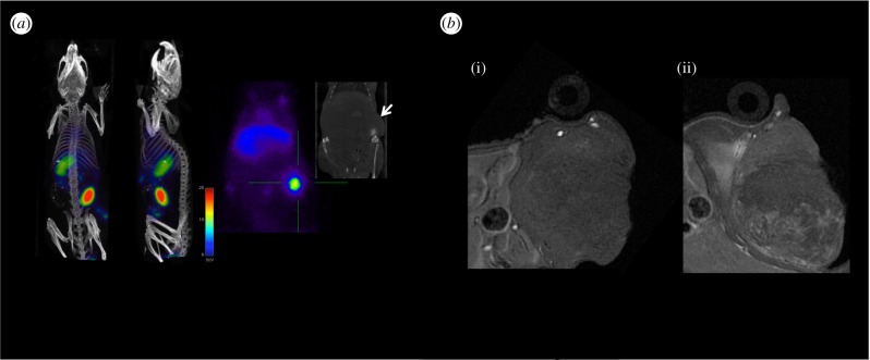
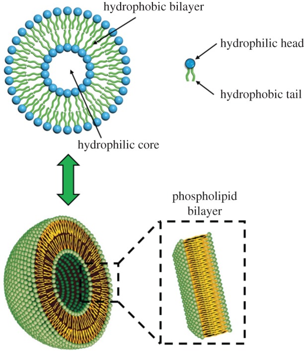
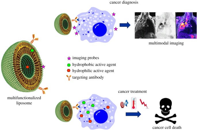
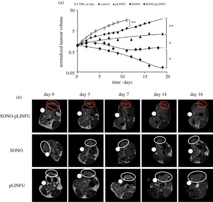
Nowadays, thanks to the successful discoveries in the biomedical field achieved in the last two decades, a deeper understanding about the complexity of mechanistic aspects of different pathological processes has been obtained. As a consequence, even the standard therapeutic protocols have undergone a vast redesign. In fact, the awareness about the necessity to progress towards a combined multitherapy in order to potentially increase the final healing chances has become a reality. One of the crucial elements of this novel approach is that large amounts of detailed information are highly needed and in vivo imaging techniques represent one of the most powerful tools to visualize and monitor the pathological state of the patient. To this scope, due to their unique features, nanostructured materials have emerged as attractive elements for the development of multifunctional tools for diagnosis and therapy. Hence, in this review, the most recent and relevant advances achieved by applying multifunctional nanostructures in multimodal theranosis of different diseases will be discussed. In more detail, the preparation and application of single multifunctional nano-radiotracers based on iron oxides and enabling PET/MRI dual imaging will be firstly detailed. After that, especially considering their highly promising clinical potential, the preparation and application of multifunctional liposomes useful for multimodal imaging and therapy will be reviewed. In both cases, a special focus will be set on the application of such a multifunctional nanocarriers in cancer as well as cardiovascular diseases.

Keywords: PET/MRI; iron oxide nanoparticles; liposome; molecular imaging; multimodal imaging; theranosis.


Figures

Figure 1.
Figure 1.
Improved overall cancer survival as a result of combination therapy. Adapted from [14].
Figure 2.
Figure 2.
Schematic of combined multitherapy in (a) cancer disease and (b) cardiovascular disease.
Figure 3.
Figure 3.
Magnetic resonance angiography of a mouse at increasing times after intravenous injection of fdIONP. Adapted from [42].
Figure 4.
Figure 4.
(a) Synthesis of 68Ga core-doped IONPs; (b) image phantoms obtained at different iron concentrations by MRI (top row) and PET (bottom row); (c) hydrodynamic size of four different samples of 68Ga core-doped IONPs; (d) thermogravimetric curve of 68Ga core-doped IONPs and (e) TEM images of the sample. Adapted from [64].
Figure 5.
Figure 5.
(a) PET/CT imaging of tumour-bearing mice 1 h post injection of 68Ga-C-IONP showing activity in tumour. (b) Axial T1-weighted MRI of tumour area in a murine model previous to injection (i) and 24 h post injection of 68Ga-C-IONP (ii). Adapted from [64].
Figure 6.
Figure 6.
General scheme of liposomes. (Online version in colour.)
Figure 7.
Figure 7.
Schematic of multifunctionalized liposome and its application for cancer diagnosis and therapy.
Figure 8.
Figure 8.
MRI analysis of the tumours after treatment with the developed multifunctionalized liposome compared with Omniscan®. (a) Increase of T1 relaxation rate (R1) over time. (b) R1 was enhanced 11- and 36-fold, respectively, 3.7 h and 4.7 h after injection with respect the commercial formulation Omniscan®. Adapted from [120].
Figure 9.
Figure 9.
MRI images of (a) tumour progression; (b) US-treated mice. The images were acquired at the time of the first MRI session (day 0) and after 3, 7, 14 and 16 days. ‘Sonoporation stimulus’ (SONO) was applied during the liposome injection, while the ‘release stimulus’ (pLINFU) was applied just after sonoporation. CTRL no LIPO refers to a group of mice that was not injected with liposomes. Adapted from [121].
Figure 10.
Figure 10.
Schematic of thermosensitive liposome encapsulating Ce6 and CuS and its working mechanism; (1) targeting delivery and cell uptake, (2) 808 nm NIR laser-induced PTT effect and PTT-induced Ce6 release and (3) 660 nm laser-induced PDT effect. Adapted from [132].

References

    1. Schork NJ. 2015. Personalized medicine: time for one-person trials. Nature 520, 609–611. ( 10.1038/520609a) - DOI - PubMed
    1. Wistuba II, Gelovani JG, Jacoby JJ, Davis SE, Herbst RS. 2011. Methodological and practical challenges for personalized cancer therapies. Nat. Rev. Clin. Oncol. 8, 135–141. ( 10.1038/nrclinonc.2011.2) - DOI - PubMed
    1. Chan IS, Ginsburg GS. 2011. Personalized medicine: progress and promise. Annu. Rev. Genomics Hum. Genet. 12, 217–244. ( 10.1146/annurev-genom-082410-101446) - DOI - PubMed
    1. Kircher MF, Hricak H, Larson SM. 2012. Molecular imaging for personalized cancer care. Mol. Oncol. 6, 182–195. ( 10.1016/j.molonc.2012.02.005) - DOI - PMC - PubMed
    1. Willmann JK, van Bruggen N, Dinkelborg LM, Gambhir SS. 2008. Molecular imaging in drug development. Nat. Rev. Drug Discov. 7, 591–607. ( 10.1038/nrd2290) - DOI - PubMed

LinkOut - more resources