Review
. 2016 Oct;2(3):136-144.
doi: 10.1159/000446336. Epub 2016 May 21.

Signaling Crosstalk between Tubular Epithelial Cells and Interstitial Fibroblasts after Kidney Injury

Affiliations
Review

Signaling Crosstalk between Tubular Epithelial Cells and Interstitial Fibroblasts after Kidney Injury

Roderick J Tan et al. Kidney Dis (Basel). 2016 Oct.

Abstract

Background: A wide variety of kidney diseases ultimately lead to tubulointerstitial damage. The initial site of injury is usually the renal tubules, with activation of fibroblasts occurring later. Self-limited disease is characterized by transient cellular activation with timed deactivation and ultimately a return to normal functioning, whereas sustained responses characterize chronic disease and the development of irreversible fibrosis. The underlying molecular and cellular mechanisms of this cascade of events remain an area of active research. Current data overwhelmingly support a role for crosstalk between the tubular epithelium and the interstitial fibroblast that mediates both repair/regeneration and progressive disease. This epithelial-mesenchymal communication (EMC) is regulated by a variety of soluble ligands binding to cell surface receptors to induce intracellular signaling events.

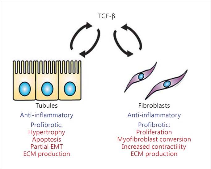
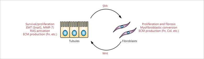
Summary: EMC is an important mechanism whereby tubular epithelium and fibroblasts/mesenchymal cells crosstalk to affect renal physiology and pathology. Numerous soluble factors such as sonic hedgehog, Wnt ligands, transforming growth factor-β, hepatocyte growth factor, connective tissue growth factor, and angiotensin II all participate in bidirectional EMC. Recent studies have also identified exosomes as a vehicle to mediate EMC during kidney injury. In general, while the short-term activity of EMC factors is renoprotective, prolonged activation of these factors leads to chronic disease and fibrosis.

Key messages: The discovery of a complex and intricate system of communication between tubular cells and fibroblasts is a new paradigm in our understanding of renal fibrosis. An appreciation of both their regenerative and pathologic functions will inform the development and use of targeted therapeutic interventions.

Keywords: Acute kidney injury; Chronic kidney disease; Inflammation; Myofibroblast; Renal fibrosis.


Figures

Fig. 1
Fig. 1
The role of TGF-β1 in EMC. Both tubules and fibroblasts can produce TGF-β1 under pathologic conditions, and this can act on either type of cell, leading to autocrine and paracrine effects. In the acute phase, TGF-β1 plays an anti-inflammatory role which may be protective. In the chronic phase, TGF-β1 is a strong inducer of fibrosis, leading to tubular cell apoptosis and hypertrophy as well as increased ECM generation and a partial EMT phenotype. In fibroblasts, TGF-β1 is a potent mitogen and stimulates myofibroblast conversion and the acquisition of a contractile phenotype along with greatly enhanced expression of ECM components.
Fig. 2
Fig. 2
Bidirectional EMC via Wnt/Shh signaling. In kidney injury, Wnt are predominantly expressed in activated proliferating fibroblasts and lead to increased β-catenin activity in tubular epithelial cells. This leads to increased cell survival as well as cell cycle progression to repair and regenerate injured tubules in the acute phase. Once the damage has been repaired, MMP-7 (itself a target of β-catenin) is secreted from tubules to induce apoptosis and return fibroblast numbers back to normal basal levels (negative feedback). However, during severe, repeated, and progressive injury, the tubules remain injured and secrete Shh, which causes profibrotic changes including further fibroblast proliferation, myofibroblast differentiation, and the increased generation of ECM. Meanwhile, these fibroblasts continue to produce Wnt, which activates β-catenin activity in tubular cells and causes EMT, ECM generation, Shh production, and upregulation of RAS (positive feedback). The resulting angiotensin II can further enhance the profibrotic changes in fibroblasts (not shown). Fn = Fibronectin; Col = collagen.
Fig. 3
Fig. 3
Shh is produced by renal tubules but acts on interstitial fibroblasts. a Immunohistochemistry for Shh after ischemia-reperfusion injury is shown with red indicating positive staining. Note the strong staining within injured tubules. b Inset showing an enlarged image of the boxed area in a. Shh staining is specific for renal tubules (black arrow) but not interstitial cells (open arrow). c Mice with the LacZ reporter under control of the Gli promoter show that only interstitial cells exhibit increased β-galactosidase activity (denoted as blue staining). d Inset showing an enlarged image of the boxed area in c. Gli reporter activity is specific for interstitial cells (black arrow) but is not present in tubular cells (open arrow).

References

    1. Levey AS, Stevens LA, Coresh J. Conceptual model of CKD: applications and implications. Am J Kidney Dis. 2009;53(suppl 3):S4–S16. - PubMed
    1. Liu ZH. Nephrology in China. Nat Rev Nephrol. 2013;9:523–528. - PubMed
    1. United States Renal Data System . 2015 USRDS annual data report: epidemiology of kidney disease in the United States. Bethesda: National Institutes of Health, National Institute of Diabetes and Digestive and Kidney Diseases; 2015.
    1. Liu Y. Cellular and molecular mechanisms of renal fibrosis. Nat Rev Nephrol. 2011;7:684–696. - PMC - PubMed
    1. Molitoris BA. Therapeutic translation in acute kidney injury: the epithelial/endothelial axis. J Clin Invest. 2014;124:2355–2363. - PMC - PubMed

LinkOut - more resources