Review
. 2017 Apr;22(2-3):253-288.
doi: 10.1007/s00775-016-1415-2. Epub 2016 Dec 5.

Activation of dioxygen by copper metalloproteins and insights from model complexes

Affiliations
Review

Activation of dioxygen by copper metalloproteins and insights from model complexes

David A Quist et al. J Biol Inorg Chem. 2017 Apr.

Abstract

Nature uses dioxygen as a key oxidant in the transformation of biomolecules. Among the enzymes that are utilized for these reactions are copper-containing metalloenzymes, which are responsible for important biological functions such as the regulation of neurotransmitters, dioxygen transport, and cellular respiration. Enzymatic and model system studies work in tandem in order to gain an understanding of the fundamental reductive activation of dioxygen by copper complexes. This review covers the most recent advancements in the structures, spectroscopy, and reaction mechanisms for dioxygen-activating copper proteins and relevant synthetic models thereof. An emphasis has also been placed on cofactor biogenesis, a fundamentally important process whereby biomolecules are post-translationally modified by the pro-enzyme active site to generate cofactors which are essential for the catalytic enzymatic reaction. Significant questions remaining in copper-ion-mediated O2-activation in copper proteins are addressed.

Keywords: Cofactor biogenesis; Copper; Copper enzymes; Dioxygen activation; Enzymatic mechanisms.


Figures

Fig. 1
Fig. 1
Synthetically derived mono- and binuclear copper-(di) oxygen complexes. Representative crystal structures are shown for the intermediates that have been structurally characterized [–, , –34]. Only the η1 superoxide and μ-η2:η2 peroxide moieties have been observed crystallographically in biological systems [–41]
Fig. 2
Fig. 2
X-ray crystal structure of the active site of PHM in its oxy-form [38]
Fig. 3
Fig. 3
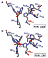
a X-ray crystal structure of LPMO with substrate bound [78]. b Catalytic reactions performed by LPMOs with products shown, derived from either H-atom abstraction at C1 or C4 [77, 80]
Fig. 4
Fig. 4
Possible reactive copper-(di)oxygen species in the catalytic cycle of LPMOs. a Tautomerization between a copper(II)-oxyl species (CuII-O·) and a copper(III) hydroxide species (CuIII-OH) [83]. b A cupric superoxide species ( CuII-O2·-) [84]
Fig. 5
Fig. 5
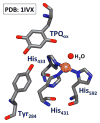
X-ray crystal structure of mature (processed) copper amine oxidase from A. globiformis in the fully oxidized form [90]
Fig. 6
Fig. 6
X-ray crystal structure of the active site of mature (processed) galactose oxidase from D. dendroides in the oxidized form [101]
Fig. 7
Fig. 7
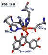
X-ray crystal structure of the enzyme-substrate complex of quercetin 2,4-dioxygenase (2,4-QD) [116]. Important hydrogen bonding between Glu73 and bound quercetin is shown as a dashed line
Fig. 8
Fig. 8
a General reaction of deoxy-Hc with O2 [131]. b X-ray crystal structure of the oxy-form of Hc from O. dofleini [130]
Fig. 9
Fig. 9
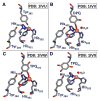
X-ray crystal structure of the a Apo-form (copper free), b Deoxy-form, c Met1-form (one water molecule), and d Oxy-form all from S. castaneoglobisporus tyrosinase [154]. This organism’s active site does not possess the His-Cys crosslink
Fig. 10
Fig. 10
X-ray crystal structure of the binuclear copper site in pMMO [186]
Fig. 11
Fig. 11
X-ray crystal structure of the resting oxidized state of the MCO, Cu-efflux oxidase (CueO) [193]. a The T1 site of MCOs. b The TNC of MCOs. c The amino acid backbone connectivity between the T1 and TNC sites
Fig. 12
Fig. 12
The four redox active metal sites of bovine heart CcO (top left) [219]. Heme a active site (bottom left). Dioxygen reduction site composed of heme a3 and CuB (bottom right). Active site of the binuclear T1 copper site referred to as CuA (top right). Bovine heart CcO numbering is used throughout the text
Fig. 13
Fig. 13
Active sites of myoglobin engineered to contain a copper binding site. Left CuBMb contains two additional histidines in the distal binding pocket [232]. Middle-Left F33Y-CuBMb and G65Y-CuBMb contain two additional histidines and a tyrosine in the distal binding pocket [235]. Middle-Right imiTyrCuBMb contains an additional histidine, and an unnatural histidine crosslinked to tyrosine in the distal binding pocket [237]. Right MII-FeBMb contains two additional histidines and a glutamate in the distal binding pocket [238]
Fig. 14
Fig. 14
Synthetic heme-copper models designed by Collman (left) [239] and Naruta (middle, right) [242, 243]
Fig. 15
Fig. 15
Synthetic heme-copper models designed by Karlin and coworkers [244, 245, 247]
Fig. 16
Fig. 16
X-ray crystal structures of a reduced [250] and b oxidized CuZnSOD [251]
Fig. 17
Fig. 17
X-ray crystal structures from A. globiformis copper amine oxidase following the biogenesis of TPQ [90]. a Preprocessed active site prior to O2 exposure. b Active site following initial tyrosine oxygenation yielding a DPQ intermediate. c Formation of TPQred following copper assisted hydrolysis of DPQ. d Processed active site following biogenesis shown in the active form
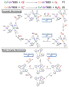
Fig. 18
Fig. 18
X-ray crystal structure of apo-GO (a) and oxidized mature GO (b) from F. graminearum [281]
Fig. 19
Fig. 19
X-ray crystal structures of a apo-Ty and b met-Ty showing the absence and formed His-Cys crosslink, respectively [284]
Scheme 1
Scheme 1
Aqueous reduction of O2 and pertinent intermediates. Reduction potentials can be found in Ref. [7]. Copper-containing enzymes that perform parts of this reduction are shown
Scheme 2
Scheme 2
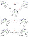
a “Flash-and-trap” technique used to photorelease CO from a CuI-CO complex in order to measure the O2 binding vs. CO rebinding kinetics in THF [48]. b Photolysis of a cupric superoxide complex, with rebinding of O2 [49]
Scheme 3
Scheme 3
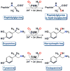
Catalytic reactions of the enzymes PHM, DβM, and TβM [57]
Scheme 4
Scheme 4
The proposed enzymatic mechanism of PHM, DβM, and TβM [17, 60, 61]
Scheme 5
Scheme 5
a [CuII(TPVMPA)(O2·-)]+ and its C–H activation reactivity [63]. b [CuII(TDMMMPA)(O2·-)]+ and its reactivity with phenolic substrates [64]. c [CuII(NDMA3S)(O2·-)]+ and its reactivity with N-methyl-9,10-dihydroacridine [67]. d [CuII(PEDACO-EtPh-R)(O2·-)]+ and its intramolecular ligand hydroxylation reactivity [52]
Scheme 6
Scheme 6
The catalytic mechanism of amine oxidase [91]. Top proposed pathway for inner-sphere reduction of O2 by CuI yielding TPQSQ and cupric superoxide [94]. Bottom proposed outer-sphere reduction of O2 by TPQAMQ yielding TPQSQ and cupric superoxide [93]
Scheme 7
Scheme 7
Transamination of benzylamine facilitated by a CuII-TPQ model complex [96]
Scheme 8
Scheme 8
The proposed catalytic mechanism of galactose oxidase [98, 104]
Scheme 9
Scheme 9
The proposed enzymatic mechanism of 2,4-QD [116]
Scheme 10
Scheme 10
a Selected example of rate enhancement from addition of exogenous acetates [123]. b Reactivity with an intramolecular acetate ligand moiety [122]. c Rate enhancement from changing electronics of the intramolecular acetate ligand moiety [124, 125]
Scheme 11
Scheme 11
Photoexcitation of a μ-η22-peroxide (top, n = 3 or 5) and a μ-1,2-peroxide (bottom) leads to the observation of novel mixed-valent dicopper superoxide complexes, which is then converted to the dicopper(I) complexes [151]. Only the starting μ-η22-peroxide fully releases O2, while the μ-1,2-peroxide keeps O2 caged until rebinding occurs
Scheme 12
Scheme 12
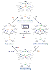
The proposed catalytic mechanism for the oxidation of o-catechol to o-quinone by catechol oxidase and tyrosinase (outer cycle), and proposed mechanism for the oxidation of phenol to o-quinone by tyrosinase (inner cycle) [12]
Scheme 13
Scheme 13
a Equilibrium between the μ-η2:η2-peroxodicopper(II) and bis(μ-oxo)dicopper(III) cores and their spectroscopic parameters [13]. b Intramolecular hydroxylation by a μ-η2:η2-peroxodicopper(II) complex [172]. c Internal hydroxylation of phenolate by a phenolate-bound bis(μ-oxo)dicopper(III) complex [171]
Scheme 14
Scheme 14
Different functions of the subclasses of coupled binuclear copper enzymes family [12]
Scheme 15
Scheme 15
The proposed electrophilic mechanism for the oxidation of o-aminophenols by NspF (hydroxyanilinase activity) [179]
Scheme 16
Scheme 16
Reduction of O2 at the TNC. O2 is first reduced by two electrons, resulting in PI which is then further reduced with cleavage of the O-O bond, resulting in NI [209]
Scheme 17
Scheme 17
Proposed catalytic mechanism of CcO [228]. See text for further explanation
Scheme 18
Scheme 18
Generation of the naked synthon through the addition of 1 equiv. of a CuI source to a ferric superoxide [248]
Scheme 19
Scheme 19
Top the two steps of superoxide dismutation by CuZnSOD. Middle the proposed enzymatic mechanism of CuZnSOD [258, 259]. Bottom the proposed mechanism for a model complex of CuZnSOD [260]
Scheme 20
Scheme 20
The proposed mechanism of TPQ or LTQ biogenesis by copper amine oxidase [87]
Scheme 21
Scheme 21
The proposed mechanism of post-synthetic modification of a CAO model compound to generate a TPQ-like moiety [96]
Scheme 22
Scheme 22
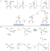
a The proposed mechanism of tyrosine-cysteine crosslink formation, starting from an initial cupric center. Both aerobic and anaerobic pathways are depicted [281]. b The proposed mechanism of tyrosine-cysteine crosslink formation, starting from an initial cuprous center [99]
Scheme 23
Scheme 23
The proposed mechanism of His-Cys formation in coupled binuclear copper enzymes [285]
Scheme 24
Scheme 24
Proposed (possible) mechanisms for an (a) O2-dependent and an (b) O2-independent formation of the Tyr-His crosslink in CcO

References

    1. Kroneck PMH, Torres MES, editors. Sustaining life on planet earth: metalloenzymes mastering dioxygen and other chewy gases. Springer International Publishing; Switzerland: 2015. - PubMed
    1. Campbell AN, Stahl SS. Acc Chem Res. 2012;45:851–863. - PMC - PubMed
    1. Mccann SD, Stahl SS. Acc Chem Res. 2015;48:1756–1766. - PubMed
    1. Gubelmann MH, Williams AF. Struct Bonding. Vol. 55. Berlin: 1983. pp. 1–65.
    1. Sawyer DT. Oxygen chemistry. Oxford University Press; New York: 1991.

Publication types