. 2016 Dec 6:6:38579.
doi: 10.1038/srep38579.

An ERβ agonist induces browning of subcutaneous abdominal fat pad in obese female mice

Affiliations

An ERβ agonist induces browning of subcutaneous abdominal fat pad in obese female mice

Yi-Fei Miao et al. Sci Rep. .

Abstract

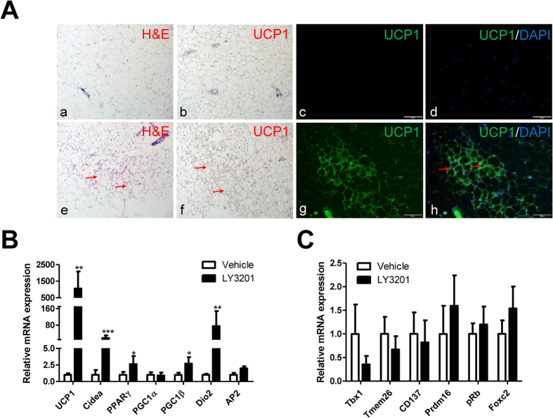
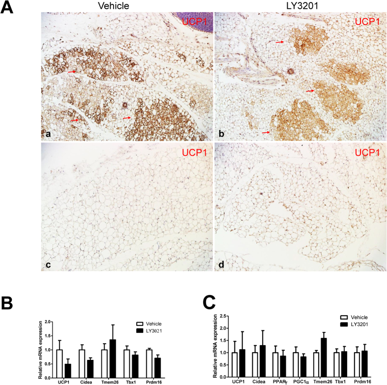
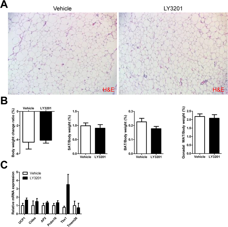
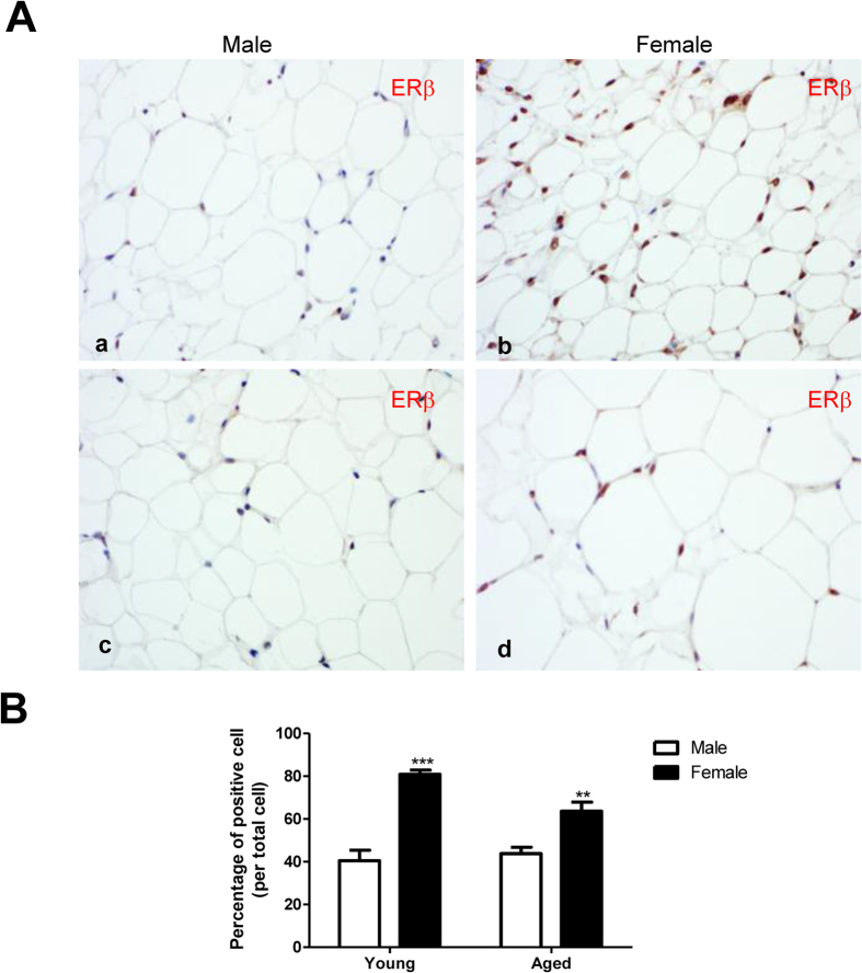
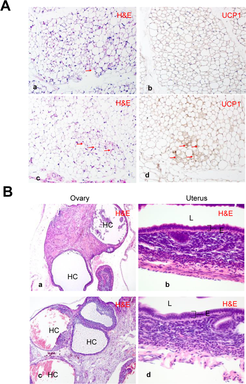
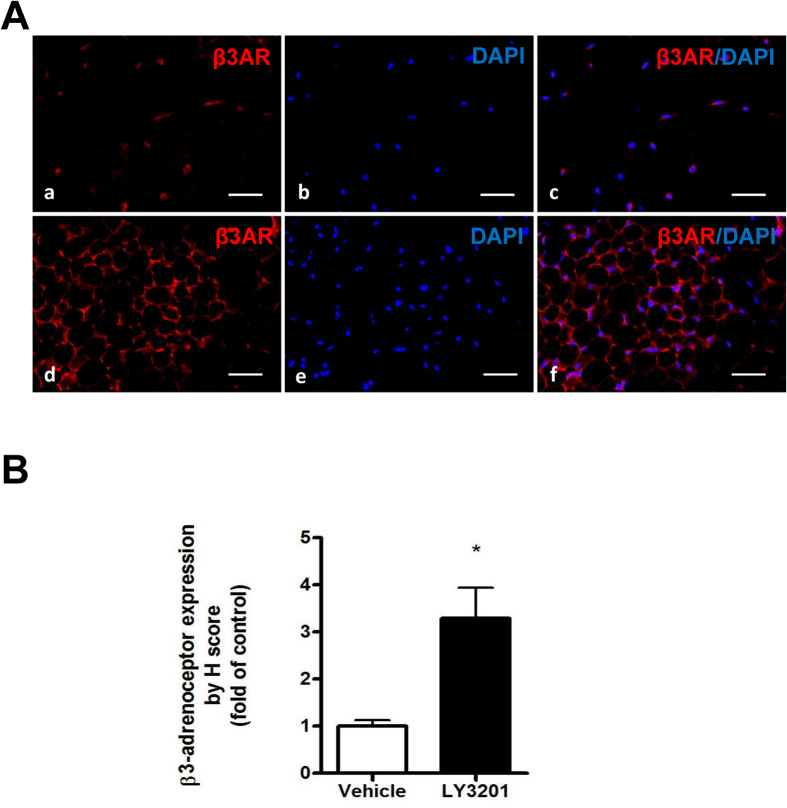
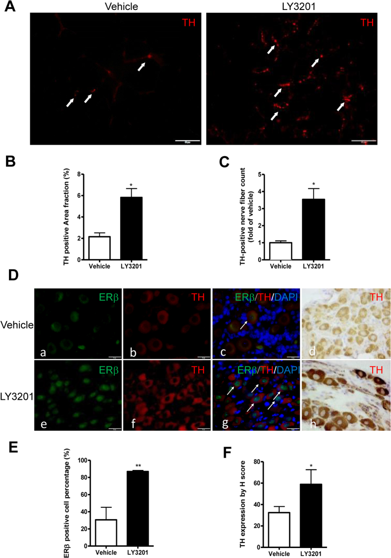
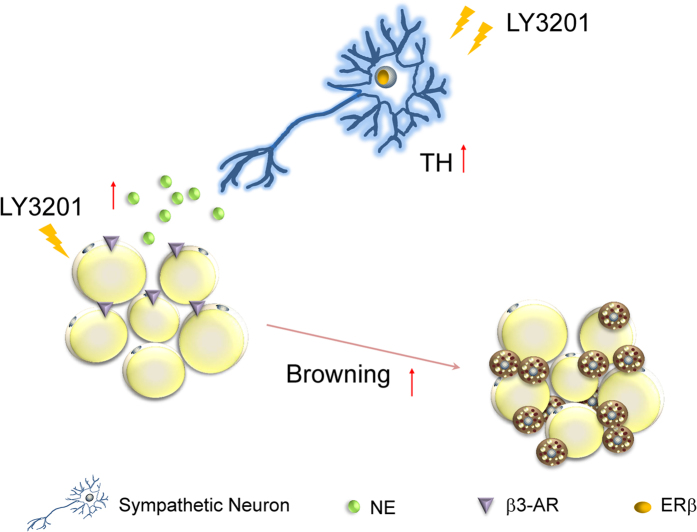
Estrogen, via estrogen receptor alpha (ERα), exerts several beneficial effects on metabolism and energy homeostasis by controlling size, enzymatic activity and hormonal content of adipose tissue. The actions of estrogen on sympathetic ganglia, which are key players in the browning process, are less well known. In the present study we show that ERβ influences browning of subcutaneous adipose tissue (SAT) via its actions both on sympathetic ganglia and on the SAT itself. A 3-day-treatment with a selective ERβ agonist, LY3201, induced browning of SAT in 1-year-old obese WT and ERα-/- female mice. Browning was associated with increased expression of ERβ in the nuclei of neurons in the sympathetic ganglia, increase in tyrosine hydroxylase in both nerve terminals in the SAT and sympathetic ganglia neurons and an increase of β3-adrenoceptor in the SAT. LY3201 had no effect on browning in young female or male mice. In the case of young females browning was already maximal while in males there was very little expression of ERβ in the SAT and very little expression of the β3-adrenoceptor. The increase in both sympathetic tone and responsiveness of adipocytes to catecholamines reveals a novel role for ERβ in controlling browning of adipose tissue.


Figures

Figure 1
Figure 1. Increased browning of SAT in obese WT female mice after treatment with LY3201 for 3 days.
(A) Treatment with LY3201 induced multilocular lipid droplets, as shown by H&E staining (red arrow) in e, and UCP1 positive (red arrow) as represented in f–h. All pictures are 20x magnification. (B) There was up-regulation of browning-associated genes by LY3201 in SAT of aged WT female mice. (C) There were no changes in beige-cell-specific genes after treatment with LY3201. *p < 0.05, **p < 0.01, ***p < 0.001, LY3201 vs. Vehicle.
Figure 2
Figure 2. Effects of 3-day treatment with LY3201 on 3-month-old female and male.
(A) UCP1 staining (red arrows) in abdominal SAT of female (a,b) or SAT of male mice (c,d). Red arrows indicate adipocytes with multilocular lipid-droplets. All pictures are 20x magnification. In line with the lack of morphological changes, LY3201 did not affect the level of mRNA of BAT-expressed genes in female (B), male (C).
Figure 3
Figure 3. Effect of LY3201 in SAT of 12-month-old male obese WT mice.
(A) LY3201 did not change the size or morphology of adipocytes in SAT as shown by H&E staining. All pictures are 20x magnification. There was no effect on body weight, adipose tissue organ weight (B) or browning or beige cell related genes (C).
Figure 4
Figure 4. Expression of ERβ in SAT from 3- and 12-month-old male and female mice.
(A) Expression of ERβ in the nuclei of SAT in young and aged male WT mice was similar (a,c). Expression of ERβ in the nuclei of SAT in 3- and 12-month-old female WT mice was higher than in age-matched male mice (b,d). (B) Quantification of ERβ. Higher expression of ERβ was observed in female SAT than in male SAT. **p < 0.01, ***p < 0.001, female vs. male. All pictures are 40x magnification.
Figure 5
Figure 5. Significant browning of abdominal SAT in aged, obese female ERα−/− mice.
(A) Treatment with LY3201 induced more multilocular lipid droplets as indicated by H&E staining (red arrow) (a,c) and expression of UCP1 (red arrow) (b,d). All pictures were 20x magnification. (B) LY3201 did not affect ovary or uterus morphology in female ERα−/− mice. As expected there were many hemorrhagic cystic follicles (HC) and lack of mature follicles and corpora lutea in ERα−/−mice. ERα−/− mice uterus was also hypoplastic with thinner epithelial layer (b,d). However, treatment with LY3201 did not further change the morphology of ovaries (a,c) or uterus (b,d). E: Epithelial layer, L: lumen of the organ. Pictures of ovaries are 10x magnification. Pictures of uterus are 20x magnification.
Figure 6
Figure 6. Expression of β3-adrenoceptor in SAT from 1-year-old obese WT female mice treated with ERβ agonist LY3201.
(A) β3-adrenoceptor (β3AR, Red) immunofluorescence in both vehicle and LY3201 treated SAT. (Blue, DAPI) (B) β3-adrenoceptor quantification in vehicle and LY3201 treated SAT. **p < 0.01, LY3201 vs. Vehicle. All pictures are 40x magnification.
Figure 7
Figure 7. Expression of TH in SAT and sympathetic ganglia from 1-year-old obese WT female mice treated with LY3201.
(A) TH expression in SAT was significantly increased after LY3201 treatment. Arrowheads indicate TH-positive fibers. TH-positive areas and TH-positive nerve fiber count in SAT in Vehicle and LY3201-treated mice were quantified (B,C). LY3201 causes a marked increase in TH expression in the SAT. (D) Immunostaining of ERβ (green) and TH (red) in thoraco-lumbar sympathetic neurons with or without LY3201 treatment (a,e,b,f). Both immunofluorescence (b,f) and immunochemistry (d,h) indicate that TH expression was strongly induced in sympathetic neurons after LY3201 treatment. Merged images of ERβ and TH (c,g). The white arrows indicate positive nuclear staining of ERβ. (E) Quantification of ERβ positive cell percentage in vehicle and LY3201-treated sympathetic ganglia. (F) TH quantification in vehicle and LY3201-treated sympathetic ganglia. All pictures are 100x magnification. *p < 0.05, LY3201 vs. Vehicle.
Figure 8
Figure 8. Schematic diagram of actions of ERβ in controlling TH expression in the sympathetic ganglia and browning of abdominal SAT in female mice with late-onset obesity.
In female mice, with late-onset obesity there is reduced ERβ expression in the nucleus of sympathetic neurons, and short term-treatment with LY3201 increased ERβ expression. LY3201 was able to up-regulate expression of TH in sympathetic neurons. Through activating the synthesis of TH, and stimulation of the expression β3-adrenoceptor LY3201 leads to the browning of SAT.

References

    1. Harms M. & Seale P. Brown and beige fat: development, function and therapeutic potential. Nat Med 19, 1252–1263, doi: 10.1038/nm.3361 (2013). - DOI - PubMed
    1. Key T. J. et al.. Body mass index, serum sex hormones, and breast cancer risk in postmenopausal women. J Natl Cancer Inst 95, 1218–1226 (2003). - PubMed
    1. Sorensen M. B., Rosenfalck A. M., Hojgaard L. & Ottesen B. Obesity and sarcopenia after menopause are reversed by sex hormone replacement therapy. Obes Res 9, 622–626, doi: 10.1038/oby.2001.81 (2001). - DOI - PubMed
    1. Heine P. A., Taylor J. A., Iwamoto G. A., Lubahn D. B. & Cooke P. S. Increased adipose tissue in male and female estrogen receptor-alpha knockout mice. Proc Natl Acad Sci USA 97, 12729–12734, doi: 10.1073/pnas.97.23.12729 (2000). - DOI - PMC - PubMed
    1. Ohlsson C. et al.. Obesity and disturbed lipoprotein profile in estrogen receptor-alpha-deficient male mice. Biochem Biophys Res Commun 278, 640–645, doi: 10.1006/bbrc.2000.3827 (2000). - DOI - PubMed

Publication types

MeSH terms