. 2017 Jan;15(1):210-220.
doi: 10.3892/mmr.2016.5979. Epub 2016 Dec 5.

Norspermidine changes the basic structure of S. mutans biofilm

Affiliations

Norspermidine changes the basic structure of S. mutans biofilm

Meizhen Ou et al. Mol Med Rep. 2017 Jan.

Abstract

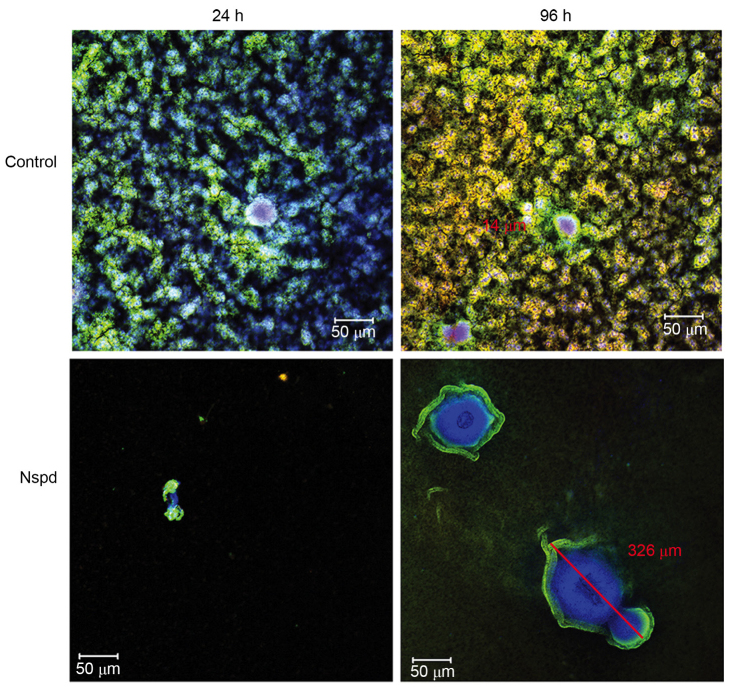
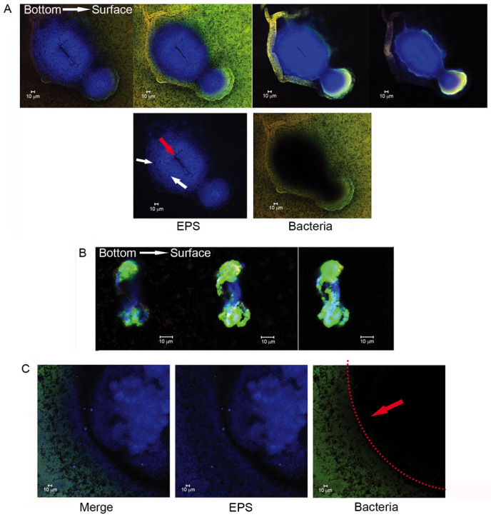
The factors regulating the assembly of the three-dimensional structure of Streptococcus mutans biofilms remain obscure. Polyamines are essential in biofilm formation of certain bacteria. Norspermidine, an unusual polyamine, has been a controversial polyamine that can lead to biofilm disassembly. However, the role of norspermidine in S. mutans biofilms remains unknown. Therefore, the present study investigated the impact of norspermidine on S. mutans biofilms. The different architectures of the biofilms in norspermidine and control groups indicated that the basic units, bacteria‑exopolysaccharide units (BEUs), represent the exopolysaccharide (EPS) and bacterial assembly pattern in S. mutans biofilms. In addition, norspermidine inhibited S. mutans biofilm formation and changed the basic composition of the biofilm, which led to an unusual EPS architecture. Therefore, 5 mM norspermidine inhibited biofilm formation both by decreasing the rate of cell viability and changing the biofilm structure. Gene‑expression microarray analysis indicated that the formation of an irregular architecture in the norspermidine group was potentially attributable to the downregulation of elements of the quorum‑sensing system (by 2.7‑15‑fold). The present study suggested that the BEUs are a basic structure of S. mutans biofilm and its assembly is regulated majorly by the quorum‑sensing system. Norspermidine can lead to structure change in BEUs by influencing S. mutans quorum-sensing system.


Figures

Figure 1.
Figure 1.
(A) Biofilms that formed following the addition of Nspd at 5 mM and incubated for 24 h, as visualized using crystal violet staining. (B) Growth curve of Streptococcus mutans. Nspd (5 mM) was added to the suspension, which was incubated anaerobically at 37°C for 12, 24, 36, 48, 60, 72, 84 and 96 h without agitation. The results were expressed as the mean ± standard deviation of triplicate assays. (C) Biofilm production was evaluated using the OD595 values. The results are expressed as the mean ± standard deviation of triplicate assays (*P<0.05, n=9 paired Student's t-test). (D) Dynamic light scattering measurements. The diameters of the water-insoluble exopolysaccharide were determined following treatment with 5 or 10 mM Nspd (n=3). Each measurement was repeated three times. Correlation analysis indicated no linear relationship between the diameter of the water-insoluble glucan and Nspd treatment (P>0.05). Nspd, norspermidine; CFU, colony forming units; OD, optical density; WIG, water-insoluble glucan.
Figure 2.
Figure 2.
Stereomicroscopic images of Streptococcus mutans biofilms that had formed by 96 h in the experimental and control groups. The biofilms were observed and images were captured using a stereomicroscope at a magnification of ×5. Nspd, norspermidine.
Figure 3.
Figure 3.
Exopolysaccharide structure of Streptococcus mutans biofilms incubated for 96 h in the experimental and control groups. Control-a, -b and -c show uniform and regular EPS granules of different areas in the control group. Nspd-a, fishnets EPS structures in the experimental group; Nspd-b, chain-like EPS structures in the experimental group; Nspd-c, star like EPS structures in the experimental group. The red arrows indicate the central hole within a polysaccharide-containing structure. The polysaccharides of the biofilms were stained blue using 300 mg/l Fluorescent Brightener 28 (magnification ×200 or ×100; scale bars, 100 µm). Nspd, norspermidine; Ctrl, control.
Figure 4.
Figure 4.
Three-dimensional distribution of exopolysaccharide and Streptococcus mutans cells in the two groups. The biofilms were incubated for 24 and 96 h, and stained using Fluorescent Brightener 28 and L-7012 to label the live (green) and dead (red) cells (scale bar, 50 µm). Nspd, norspermidine.
Figure 5.
Figure 5.
Bacteria-containing ‘hollow sphere’ structures in the Streptococcus mutans biofilms. The images are sections of 3D imaging demonstrating representative images from the bottom, middle and surface of the biofilms. The biofilms were formed in brain heart infusion broth containing 1% sucrose during a 12 h incubation and were stained using the LIVE/DEAD kit to label the live (green) and dead (red) cells. The red arrows indicate a ‘space’ in a spherical structure (magnification ×200; scale bar, 10 µm).
Figure 6.
Figure 6.
Normal BEUs in Streptococcus mutans biofilms. The images in the Merge row reveal the EPS and bacteria. Images in the EPS row highlight the EPS with the units and the images in the Bacteria row show the bacteria on the exterior of the units. The red arrows indicate typical BEUs consisting of bacteria on the exterior and EPS in the interior. (A) A single BEU with bacteria surrounding an EPS pearl. (B) A combination of two BEUs. The white arrow indicates a typical toroid combined-BEU structure. (C) A single BEU with bacteria surrounding the EPS. The white arrow indicates a rough chain-like combined-BEU structure. The biofilms were stained using 300 mg/l Fluorescent Brightener 28 to label the polysaccharides (blue), and a L-7012 LIVE/DEAD Viability kit to label the live (green) and dead (red) cells (scale bar, 10 µm). EPS, exopolysaccharide; BEUs, bacteria-EPS units.
Figure 7.
Figure 7.
HugeBEUs in the Streptococcus mutans biofilms of the Nspd group. (A) Typical large-scale mature BEUs. The first panel shows optical sections of a unit starting at the adherent interface (bottom) and moving to the upper surface of biofilm surrounded by liquid. The second panel shows detailed views of separate and merged images of the EPS and bacterial cells. (B) Optical sections showing initially formed irregular BEUs, starting at the bottom and moving to the surface. These images show that the bacterial cells secreted EPS toward the center of the units and the bottom interface. (C) A typical spherical interface between the bacteria and EPS of developing BEUs. This image shows the cellular arrangement and the EPS-secretion pattern. The biofilms were stained using 300 mgl Fluorescent Brightener 28 to label the polysaccharides (blue), and a L-7012 LIVE/DEAD Viability kit to label the live (green) and dead (red) cells. The red arrows indicates typical basic units consisting of bacteria on the exterior and EPS in the interior (scale bar, 10 µm). EPS, exopolysaccharide; BEUs, bacteria-EPS units.
Figure 8.
Figure 8.
Overview of the Streptococcus mutans genes and pathways regulated by Nspd. (A) Volcano plots to visualize the differential gene expression in the Nspd group compared with the control group. The vertical lines correspond to a 1.5-fold difference in expression levels, and the horizontal line represents a P=0.05. The red dots in the plot represent mRNAs that were differentially expressed at significant levels. (B) Pathways that were upregulated in the Nspd group compared with the control group. (C) Pathways that were downregulated in the Nspd group compared with the control group. These data were obtained using the latest Kyoto Encyclopedia of Genes and Genomes database. The name of the pathway is noted on the left. The P-value indicates the significance of differential expression of the genes in the pathway. The smaller the P-value, the more significantly differentially expressed were the genes in the pathway (P=0.05 was the cut-off). The P-value was calculated using Fisher's exact test. The enrichment scores were expressed as -log10 units (P-value). Nspd, norspermidine; Sig, signaling; DE, differentially expressed.
Figure 9.
Figure 9.
Network map of biofilm-associated genes, QS-associated genes and polyamine-associated genes. (A) The relationship among the polyamine-synthetase genes and the luxS gene. (B) The regulation of the biofilm-associated genes by the competence QS genes and AI-2 QS genes. This figure was created by our research group using the program at http://string-db.org/newstring.
Figure 10.
Figure 10.
A schematic drawing of the three stages of Streptococcus mutans biofilm formation via BEUs in the control, Spd and Nspd groups. Nspd, norspermidine; BEU, bacteria-exopolysaccharide units.

References

    1. Wallace HM, Fraser AV, Hughes A. A perspective of polyamine metabolism. Biochem J. 2003;376:1–14. doi: 10.1042/bj20031327. - DOI - PMC - PubMed
    1. Romero D, Kolter R. Will biofilm disassembly agents make it to market? Trends Microbiol. 2011;19:304–306. doi: 10.1016/j.tim.2011.03.003. - DOI - PMC - PubMed
    1. Flemming HC, Neu TR, Wozniak DJ. The EPS matrix: The ‘house of biofilm cells’. J Bacteriol. 2007;189:7945–7947. doi: 10.1128/JB.00858-07. - DOI - PMC - PubMed
    1. Flemming HC, Wingender J. The biofilm matrix. Nat Rev Microbiol. 2010;8:623–633. - PubMed
    1. Branda SS, Vik S, Friedman L, Kolter R. Biofilms: The matrix revisited. Trends Microbiol. 2005;13:20–26. doi: 10.1016/j.tim.2004.11.006. - DOI - PubMed

MeSH terms