. 2016 Dec 4;8(12):3375-3389.
doi: 10.18632/aging.101126.

Impaired energy metabolism of senescent muscle satellite cells is associated with oxidative modifications of glycolytic enzymes

Affiliations

Impaired energy metabolism of senescent muscle satellite cells is associated with oxidative modifications of glycolytic enzymes

Martín A Baraibar et al. Aging (Albany NY). .

Abstract

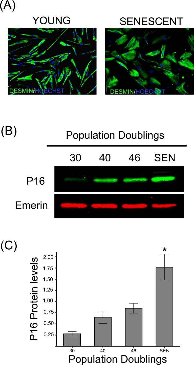
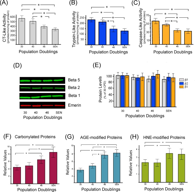
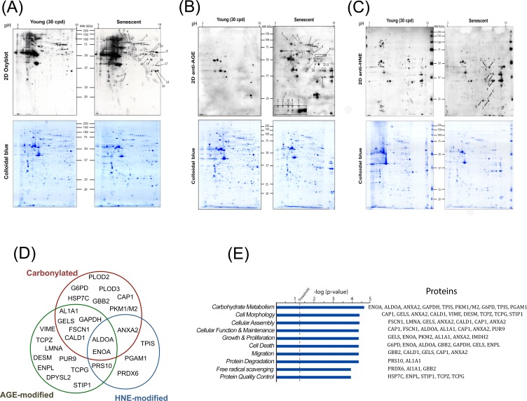
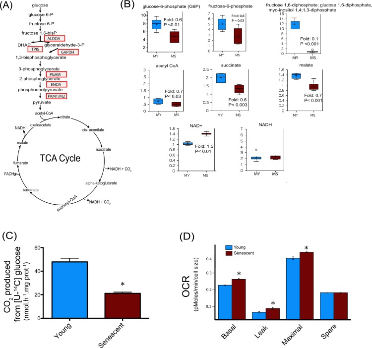
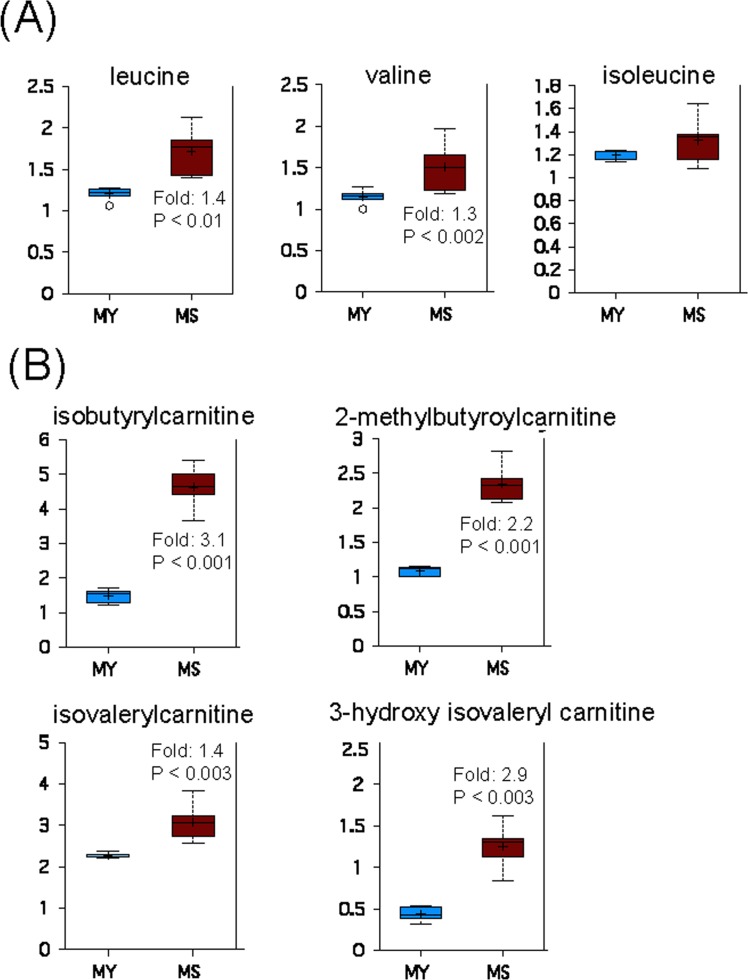
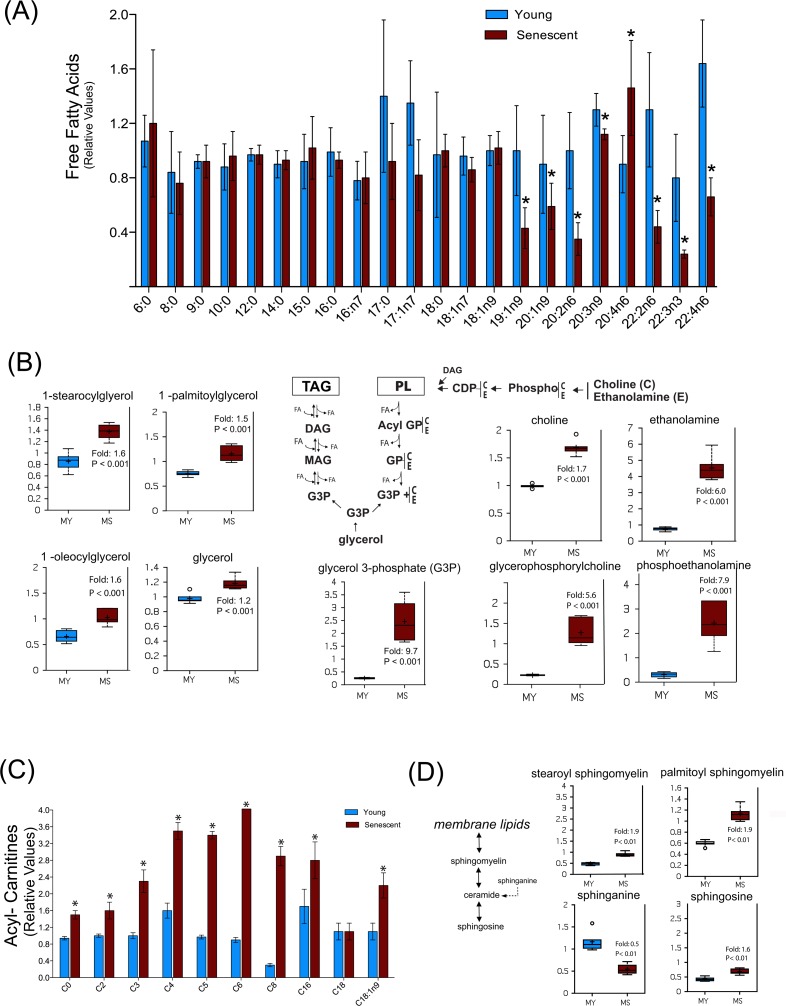
Accumulation of oxidized proteins is a hallmark of cellular and organismal aging. Adult muscle stem cell (or satellite cell) replication and differentiation is compromised with age contributing to sarcopenia. However, the molecular events related to satellite cell dysfunction during aging are not completely understood. In the present study we have addressed the potential impact of oxidatively modified proteins on the altered metabolism of senescent human satellite cells. By using a modified proteomics analysis we have found that proteins involved in protein quality control and glycolytic enzymes are the main targets of oxidation (carbonylation) and modification with advanced glycation/lipid peroxidation end products during the replicative senescence of satellite cells. Inactivation of the proteasome appeared to be a likely contributor to the accumulation of such damaged proteins. Metabolic and functional analyses revealed an impaired glucose metabolism in senescent cells. A metabolic shift leading to increased mobilization of non-carbohydrate substrates such as branched chain amino acids or long chain fatty acids was observed. Increased levels of acyl-carnitines indicated an increased turnover of storage and membrane lipids for energy production. Taken together, these results support a link between oxidative protein modifications and the altered cellular metabolism associated with the senescent phenotype of human myoblasts.

Keywords: aging; cellular senescence; energy metabolism; myoblasts; protein oxidation; proteostasis.


Conflict of interest statement

The authors have no conflict of interests to declare.

Figures

Figure 1
Figure 1. Replicative senescence of human satellite cells in vitro
(A) Immunocytochemistry against desmin evidences morphological changes in senescent human myoblasts (SEN) when compared to young cells (CPD 30). Note the increased diameter and irregular shapes of the formers, as previously described [13]. (B) p16 protein levels during replicative senescence in human myoblasts. (C) Densitometric analysis of the p16/emerin ratio showed a significant increase (n=3; *P<0.001) in p16 levels during replicative senescence. P16 protein levels are expressed as relative values and shown as mean ± S.D.
Figure 2
Figure 2. Decreased proteasome activity is associated with the accumulation of oxidized and damaged proteins during replicative senescence
Chymotrypsin like (A), trypsin-like (B), and caspase-like (C) peptidase activities of the proteasome were measured during replicative senescence. Protein levels of proteasome catalytic subunits (β1, β2, and β5) were assessed by western blot (D) and catalytic subunits protein levels were quantified by densitometric analysis (E). Quantification of carbonylated proteins (F), proteins modified by different glycated end products (G), or modified by the lipid peroxidation product 4-hydroxynonenal (H) during replicative senescence of human myoblasts. Protein modifications are expressed as relative values and shown as mean±S.D. (n=3). Data were analyzed by two-way ANOVA followed by Bonferroni's post hoc test. * P<0.05.
Figure 3
Figure 3. Identification and data mining of modified proteins
Cellular protein extracts from young (30 CPD) and senescent human myoblasts were separated by two-dimensional gel electrophoresis. After the second dimension, gels were either stained with colloidal Coomassie Brilliant Blue G (bottom panels) or electrotransferred onto nitrocellulose membranes for subsequently detection of: carbonylated proteins using the OxyBlotTM kit (A); glycoxidation protein adducts (B) and HNE-modified proteins (C). Presented results are from one representative experiment of three independent experiments using three different batches of cells. Numbers refer to the spots evidenced as consistently increased in senescent cells identified by MS/MS. (D) Venn diagram depicting the distribution of proteins in relation with the modifications studied (see also Table S1, Table S2 and Table S3). (E) Modified proteins were grouped into functional categories through the use of Ingenuity Pathways Analysis. The bars represent the biological functions identified, named in the x-axis. The dotted line represents the threshold above which there are statistically significantly more proteins in a biological function than expected by chance. The identified proteins associated with each pathway are indicated.
Figure 4
Figure 4. Central metabolism alterations in senescent satellite cells
(A) Modified enzymes identified in senescent cells and related to the central metabolism are represented in boxes. (B) Altered metabolites of central metabolism profiling in young (30 CPD) (MY) and senescent myoblast (MS). For the box plots, the top and bottom of the boxes represent the 75th and 25th percentile, respectively. The solid bar across the box represents the median value, while the + is the mean. Any statistical outliers are represented by a circle. The Y axis is the median scaled value (relative level). The fold change and the corresponding p value in senescent cells relative to their young counterpart is indicated in each plot (see also Data set 1). (C) Glucose flux in young and senescent myoblasts measured by [U-14C] glucose oxidation into 14CO2. (D) Oxygen consumption rates (OCR) of young and senescent myoblasts were monitored using the Seahorse Bioscience Extra Cellular Flux Analyzer. Mitochondrial respiration was determined in basal conditions (growth media), in the presence of oligomycin (leak), and finally in the presence of increasing amounts of carbonyl cyanide m-chlorophenylhydrazone (CCCP; 1-30 μM) to determine the maximal respiration rate. The respiration reserve capacity (spare) was calculated by subtracting the basal to the maximal respiration. The OCR values were normalized to cellular size.
Figure 5
Figure 5. Catabolism of BCAAs is increased during replicative senescence
(A) Relative levels of branched chain amino acids in young (MY) and senescent (MS) myoblasts. (B) Carnitine conjugates of BCAA-derived biochemicals. For details of box plots see Figure legend 4.
Figure 6
Figure 6. Senescent satellite cells exhibit altered lipid metabolism
(A) Free fatty acids profiling in young (MY) and senescent myoblasts (MS). (B) Increased glycerolipids turnover in senescent cells. PL: phospholipids; TAG: triacylglycerols; DAG: diacylglycerols; MAG: monoacylglycerols; FA: fatty acids; GP: glycerol phosphate. C: choline; E: ethanolamine. (C) Free carnitine and acylcarnitine profiling. The acyl chain length (c) is denoted by the corresponding metabolite number (e.g., C0 = free carnitine, C2 = acetylcarnitine; C3 =proprionylcarnitine). (D) Sphingolipids metabolism in young and senescent myoblasts. Data are expressed as mean ± S.E.M of six independent experiments. * p <0.05. For details of box plots see Figure legend 4.

References

    1. Cruz-Jentoft AJ, Baeyens JP, Bauer JM, Boirie Y, Cederholm T, Landi F, Martin FC, Michel JP, Rolland Y, Schneider SM, Topinková E, Vandewoude M, Zamboni M. and European Working Group on Sarcopenia in Older People. Sarcopenia: European consensus on definition and diagnosis: Report of the European Working Group on Sarcopenia in Older People. Age Ageing. 2010;39:412–23. doi: 10.1093/ageing/afq034. - DOI - PMC - PubMed
    1. Lepper C, Partridge TA, Fan CM. An absolute requirement for Pax7-positive satellite cells in acute injury-induced skeletal muscle regeneration. Development. 2011;138:3639–46. doi: 10.1242/dev.067595. - DOI - PMC - PubMed
    1. Mauro A. Satellite cell of skeletal muscle fibers. J Biophys Biochem Cytol. 1961;9:493–95. doi: 10.1083/jcb.9.2.493. - DOI - PMC - PubMed
    1. Chargé SB, Rudnicki MA. Cellular and molecular regulation of muscle regeneration. Physiol Rev. 2004;84:209–38. doi: 10.1152/physrev.00019.2003. - DOI - PubMed
    1. Renault V, Thornell LE, Butler-Browne G, Mouly V. Human skeletal muscle satellite cells: aging, oxidative stress and the mitotic clock. Exp Gerontol. 2002;37:1229–36. doi: 10.1016/S0531-5565(02)00129-8. - DOI - PubMed

Publication types