. 2016 Dec 6;12(12):e1006461.
doi: 10.1371/journal.pgen.1006461. eCollection 2016 Dec.

Mutations in the Heme Exporter FLVCR1 Cause Sensory Neurodegeneration with Loss of Pain Perception

Affiliations

Mutations in the Heme Exporter FLVCR1 Cause Sensory Neurodegeneration with Loss of Pain Perception

Deborah Chiabrando et al. PLoS Genet. .

Abstract

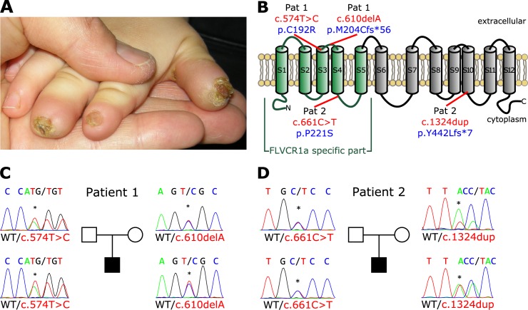
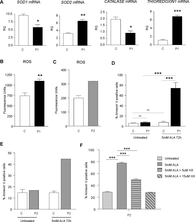
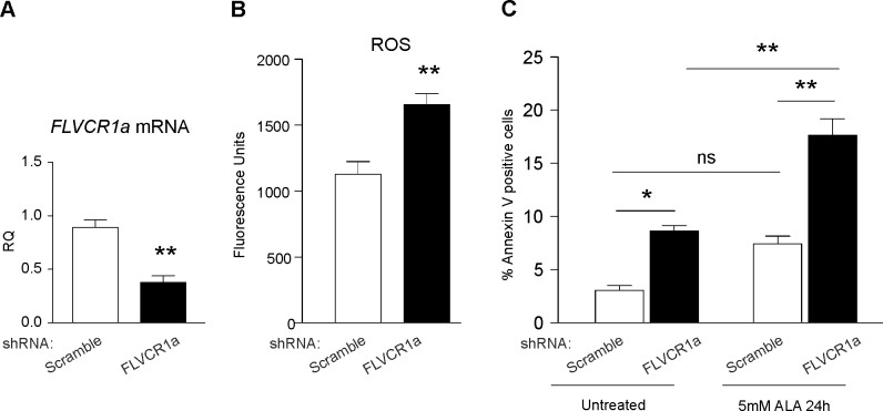
Pain is necessary to alert us to actual or potential tissue damage. Specialized nerve cells in the body periphery, so called nociceptors, are fundamental to mediate pain perception and humans without pain perception are at permanent risk for injuries, burns and mutilations. Pain insensitivity can be caused by sensory neurodegeneration which is a hallmark of hereditary sensory and autonomic neuropathies (HSANs). Although mutations in several genes were previously associated with sensory neurodegeneration, the etiology of many cases remains unknown. Using next generation sequencing in patients with congenital loss of pain perception, we here identify bi-allelic mutations in the FLVCR1 (Feline Leukemia Virus subgroup C Receptor 1) gene, which encodes a broadly expressed heme exporter. Different FLVCR1 isoforms control the size of the cytosolic heme pool required to sustain metabolic activity of different cell types. Mutations in FLVCR1 have previously been linked to vision impairment and posterior column ataxia in humans, but not to HSAN. Using fibroblasts and lymphoblastoid cell lines from patients with sensory neurodegeneration, we here show that the FLVCR1-mutations reduce heme export activity, enhance oxidative stress and increase sensitivity to programmed cell death. Our data link heme metabolism to sensory neuron maintenance and suggest that intracellular heme overload causes early-onset degeneration of pain-sensing neurons in humans.


Conflict of interest statement

The authors have declared that no competing interests exist.

Figures

Fig 1
Fig 1. Mutations in the FLVCR1 gene cause Sensory Neuropathy.
(A) Non-healing wound lesions with nail dystrophy in patient 2 as clinical feature of sensory neuropathy caused by FLVCR1 mutations. (B) Structure of the FLVCR1a protein (plasma membrane isoform). FLVCR1a-specific part is depicted in green. Mutations are indicated in the scheme, electropherograms with mutations and pedigrees are given for patient 1 (C) and patient 2 (D).
Fig 2
Fig 2. FLVCR1 mutations impair heme export in patient-derived cells.
(A) qRT-PCR analysis of FLVCR1a mRNA in patient 1 compared to control fibroblasts (black). Values represent mean ± SEM. n = 6. * = P<0.05. (B) qRT-PCR analysis of FLVCR1a mRNA in patient 2 compared to control LCLs (grey). Values represent mean FLVCR1a mRNA levels in patient 2 compared to the mean FLVCR1a mRNA levels of 4 different control LCLs. (C) Immunoprecipitation and western blotting of FLVCR1a in patient 1 compared to control fibroblasts. A representative blot is shown. The antibody against SUMO was used as control. (D) Immunoprecipitation and western blotting of FLVCR1a in patient 2 compared to control LCLs. A representative blot is shown. The antibody against SUMO was used as control. (E) qRT-PCR analysis of ALAS1, HO1, FT-L, FT-H and FPN1 mRNA in patient compared to control fibroblasts. Values represent mean ± SEM. n = 6. * = P<0.05; *** = P<0.001. (F) qRT-PCR analysis of ALAS1, HO1, FT-L, FT-H and FPN1 mRNA in patient compared to control LCLs. Values represent mean mRNA levels in patient 2 compared to the mean mRNA levels of 4 different control LCLs. (G) Western blot analysis of HO1 and ALAS1 protein in patient 1 compared to control fibroblasts. A representative blot is shown. (H) Western blot analysis of ALAS1 and HO1 protein levels in patient 2 compared to 4 different control LCLs. A representative blot is shown. (I) Measurement of heme content in patient 1 compared to control fibroblasts. Values represent mean ± SEM. n = 6. Two-way ANOVA. *** = P<0.001. (J) Measurement of heme content in patient 1 compared to control LCLs. Values represent heme content of patient 2 LCLs compared to the mean heme content of 4 different control LCLs. P1 = patient 1, P2 = patient 2, C = control.
Fig 3
Fig 3. FLVCR1 mutations induce oxidative stress and increase the sensitivity to programmed cell death in patient-derived cells.
(A) qRT-PCR analysis of SOD1, SOD2, CATALASE and THIOREDOXIN1 mRNAs in patient 1 compared to control fibroblasts. Values represent mean ± SEM. n = 3. * = P<0.05; ** = P<0.005; *** = P<0.001. (B) Measurement of ROS levels in patient 1 compared to control fibroblasts. Values represent mean ± SEM. n = 6. ** = P<0.005. (C) Measurement of ROS levels in patient 2 compared to control LCLs. Values represent ROS levels of patient 2 LCLs compared to the mean heme content of 4 different control LCLs. (D) Percentage of Annexin V-positive cells in patient 1 compared to control fibroblasts, under basal conditions and following the stimulation with 5mM ALA for 72 hours. Values represent mean ± SEM. n = 6. Two-way ANOVA. *** = P<0.001. (E) Annexin V-positive cells in patient 2 compared to control LCLs under basal conditions and following the stimulation with 5mM ALA for 72 hours. Values represent the percentage of Annexin V-positive cells of patient 2 LCLs compared to the mean percentage of Annexin V-positive cells of 4 different control LCLs. (F) Annexin V-positive cells in patient LCLs grown in starved medium, treated with 5mM ALA and with or without Hemopexin (HX) for 24 hours. Values represent mean ± SEM. n = 6. Two-way ANOVA. *** = P<0.001. P1 = patient 1, P2 = patient 2, C = control.
Fig 4
Fig 4. FLVCR1a is important for the survival of neuroblastoma cells.
(A) qRT-PCR analysis of FLVCR1a mRNA levels in FLVCR1a-depleted SH-SY5Y cells compared to control (scramble). Values represent mean ± SEM. n = 3. ** = P<0.005. (B) Measurement of ROS levels mRNA in FLVCR1a-downregulated SH-SY5Y cells compared to controls. Values represent mean ± SEM. n = 6. ** = P<0.005. (C) Annexin V staining of FLVCR1a-downregulated SH-SY5Y cells compared to controls. Values represent mean ± SEM. n = 3. Two-way ANOVA. * = P<0.05; ** = P<0.005.

References

    1. Rotthier A, Baets J, Timmerman V, Janssens K. Mechanisms of disease in hereditary sensory and autonomic neuropathies. Nat Rev Neurol. 2012;8(2):73–85. 10.1038/nrneurol.2011.227 - DOI - PubMed
    1. Auer-Grumbach M. Hereditary sensory and autonomic neuropathies. Handb Clin Neurol. 2013;115:893–906. 10.1016/B978-0-444-52902-2.00050-3 - DOI - PubMed
    1. Bennett DL, Woods CG. Painful and painless channelopathies. Lancet Neurol. 2014;13(6):587–99. 10.1016/S1474-4422(14)70024-9 - DOI - PubMed
    1. Verpoorten N, De Jonghe P, Timmerman V. Disease mechanisms in hereditary sensory and autonomic neuropathies. Neurobiol Dis. 2006;21(2):247–55. 10.1016/j.nbd.2005.08.004 - DOI - PubMed
    1. Khaminets A, Heinrich T, Mari M, Grumati P, Huebner AK, Akutsu M, et al. Regulation of endoplasmic reticulum turnover by selective autophagy. Nature. 2015;522(7556):354–8. 10.1038/nature14498 - DOI - PubMed

MeSH terms