Case Reports
. 2016 Dec 7;16(1):213.
doi: 10.1186/s12886-016-0394-y.

Postoperative occlusion of visual axis with fibrous membrane in the presence of anterior capsular phimosis in a patient with pseudoexfoliation syndrome: a case report

Affiliations
Case Reports

Postoperative occlusion of visual axis with fibrous membrane in the presence of anterior capsular phimosis in a patient with pseudoexfoliation syndrome: a case report

Eung Suk Kim et al. BMC Ophthalmol. .

Abstract

Background: To report a case of postoperative fibrous membrane formation occluding the visual axis in the presence of anterior capsular phimosis in a patient with pseudoexfoliation syndrome.

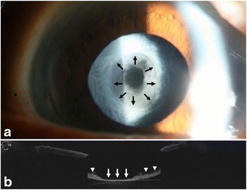
Case presentation: A 79-year-old Asian woman with pseudoexfoliation syndrome underwent uneventful phacoemulsification and implantation of one-piece hydrophilic acrylic square-edged intraocular lens (Cristalens) in the right eye. Two months later, she had blurred vision in the right eye with the best-corrected visual acuity (BCVA) of 20/40. Formation of fibrous membrane occluding the capsulorhexis opening with contraction of anterior capsule was observed, which was confirmed by anterior segment optical coherence tomography. Clear visual axis was achieved by lysis of the membrane using Nd:YAG laser. The BCVA improved to 20/20.

Conclusions: Occlusion of the visual axis with fibrous membrane can develop in the presence of anterior capsular phimosis in a patient with pseudoexfoliation syndrome.

Keywords: Anterior capsular phimosis; Anterior segment optical coherence tomography; Case report; Fibrous membrane; Nd:YAG Laser; Pseudoexfoliation syndrome.


Figures

Fig. 1
Fig. 1
Anterior segment examnation of the right eye shows shrinkage of the anterior capsulorhexis opening and fibrous membrane formation occluding the opening. Margin of the anterior capsulorhexis is clearly visible (black arrow) (a). An AS-OCT image shows the presence of fibrous membrane occluding the visual axis (white arrow) and thick anterior capsule (white arrowhead) (b)
Fig. 2
Fig. 2
Immediately after the Nd:YAG laser treatment, anterior segment exmination of the right eye demonsrtates a clear anterior capsular opening (a). An AS-OCT image confirms removal of the fibrous membrane (white arrow) surrounded by thick anterior capsule (white arrowhead) (b)
Fig. 3
Fig. 3
At 1 month after the Nd:YAG laser treatment, anterior segment examination of the right eye reveals clear visual axis (a). An AS-OCT image shows clear anterior surface of the IOL (white arrow) (b)

References

    1. Davison JA. Capsule contraction syndrome. J Cataract Refract Surg. 1993;19:582–9. doi: 10.1016/S0886-3350(13)80004-1. - DOI - PubMed
    1. Spang KM, Rohrbach JM, Weidle EG. Complete occlusion of the anterior capsular opening after intact capsulorhexis: clinicopathologic correlation. Am J Ophthalmol. 1999;127:343–5. doi: 10.1016/S0002-9394(98)00323-7. - DOI - PubMed
    1. Moreno-Montanes J, Sanchez-Tocino H, Rodriguez-Conde R. Complete anterior capsule contraction after phacoemulsification with acrylic intraocular lens and endocapsular ring implantation. J Cataract Refract Surg. 2002;28:717–9. doi: 10.1016/S0886-3350(01)01231-7. - DOI - PubMed
    1. Zaugg B, Werner L, Neuhann T, et al. Clinicopathologic correlation of capsulorhexis phimosis with anterior flexing of single-piece hydrophilic acrylic intraocular lens haptics. J Cataract Refract Surg. 2010;36:1605–9. doi: 10.1016/j.jcrs.2010.06.029. - DOI - PubMed
    1. Dubois VD, Ainsworth G, Liu CS. Unilateral capsular phimosis with an acrylic IOL and two capsular tension rings in pseudoexfoliation. Clin Experiment Ophthalmol. 2009;37:631–3. doi: 10.1111/j.1442-9071.2009.02051.x. - DOI - PubMed

Publication types