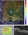
Retinal microvascular network alterations: potential biomarkers of cerebrovascular and neural diseases
- PMID: 27923786
- PMCID: PMC5336575
- DOI: 10.1152/ajpheart.00201.2016
Retinal microvascular network alterations: potential biomarkers of cerebrovascular and neural diseases
Abstract
Increasing evidence suggests that the conditions of retinal microvessels are indicators to a variety of cerebrovascular, neurodegenerative, psychiatric, and developmental diseases. Thus noninvasive visualization of the human retinal microcirculation offers an exceptional opportunity for the investigation of not only the retinal but also cerebral microvasculature. In this review, we show how the conditions of the retinal microvessels could be used to assess the conditions of brain microvessels because the microvascular network of the retina and brain share, in many aspects, standard features in development, morphology, function, and pathophysiology. Recent techniques and imaging modalities, such as optical coherence tomography (OCT), allow more precise visualization of various layers of the retina and its microcirculation, providing a "microscope" to brain microvessels. We also review the potential role of retinal microvessels in the risk identification of cerebrovascular and neurodegenerative diseases. The association between vision problems and cerebrovascular and neurodegenerative diseases, as well as the possible role of retinal microvascular imaging biomarkers in cerebrovascular and neurodegenerative screening, their potentials, and limitations, are also discussed.
Keywords: brain; cerebrovascular and neurodegenerative disorders; optical coherence tomography; optical imaging; retinal imaging biomarkers.
Copyright © 2017 the American Physiological Society.
Figures


Similar articles
-
Correlation between corneal and retinal neurodegenerative changes and their association with microvascular perfusion in type II diabetes.Acta Ophthalmol. 2019 Jun;97(4):e545-e550. doi: 10.1111/aos.13938. Epub 2018 Oct 11. Acta Ophthalmol. 2019. PMID: 30311432
-
Imaging retinal microvascular manifestations of carotid artery disease in older adults: from diagnosis of ocular complications to understanding microvascular contributions to cognitive impairment.Geroscience. 2021 Aug;43(4):1703-1723. doi: 10.1007/s11357-021-00392-4. Epub 2021 Jun 8. Geroscience. 2021. PMID: 34100219 Free PMC article. Review.
-
Optical coherence tomography angiography analysis of retinal vascular plexuses and choriocapillaris in patients with type 1 diabetes without diabetic retinopathy.Acta Diabetol. 2017 Jul;54(7):695-702. doi: 10.1007/s00592-017-0996-8. Epub 2017 May 5. Acta Diabetol. 2017. PMID: 28474119
-
Assessment of superficial retinal microvascular density in healthy myopia.Int Ophthalmol. 2019 Aug;39(8):1861-1870. doi: 10.1007/s10792-018-1014-z. Epub 2018 Sep 3. Int Ophthalmol. 2019. PMID: 30178185
-
Retinal Vascular Signs and Cerebrovascular Diseases.J Neuroophthalmol. 2020 Mar;40(1):44-59. doi: 10.1097/WNO.0000000000000888. J Neuroophthalmol. 2020. PMID: 31977663 Review.
Cited by
-
Association between functional network connectivity, retina structure and microvasculature, and visual performance in patients after thalamic stroke: An exploratory multi-modality study.Brain Behav. 2024 Jan;14(1):e3385. doi: 10.1002/brb3.3385. Brain Behav. 2024. PMID: 38376035 Free PMC article.
-
Ganglion Cell Layer Thinning in Alzheimer's Disease.Medicina (Kaunas). 2020 Oct 21;56(10):553. doi: 10.3390/medicina56100553. Medicina (Kaunas). 2020. PMID: 33096909 Free PMC article. Review.
-
Predicting the severity of white matter lesions among patients with cerebrovascular risk factors based on retinal images and clinical laboratory data: a deep learning study.Front Neurol. 2023 Jul 10;14:1168836. doi: 10.3389/fneur.2023.1168836. eCollection 2023. Front Neurol. 2023. PMID: 37492851 Free PMC article.
-
Association of Retinal Microvascular Characteristics With Short-term Memory Performance in Children Aged 4 to 5 Years.JAMA Netw Open. 2020 Jul 1;3(7):e2011537. doi: 10.1001/jamanetworkopen.2020.11537. JAMA Netw Open. 2020. PMID: 32706383 Free PMC article.
-
Ophthalmic characteristics and retinal vasculature changes in Williams syndrome, and its association with systemic diseases.Eye (Lond). 2023 Aug;37(11):2265-2271. doi: 10.1038/s41433-022-02328-4. Epub 2022 Nov 28. Eye (Lond). 2023. PMID: 36437422 Free PMC article.
References
-
- Baker ML, Hand PJ, Liew G, Wong TY, Rochtchina E, Mitchell P, Lindley RI, Hankey GJ, Wang JJ; Multi-Centre Retinal Stroke Study Group . Retinal microvascular signs may provide clues to the underlying vasculopathy in patients with deep intracerebral hemorrhage. Stroke 41: 618–623, 2010. doi:10.1161/STROKEAHA.109.569764. - DOI - PubMed
Publication types
MeSH terms
LinkOut - more resources
Full Text Sources
Other Literature Sources
Medical

