. 2017 Mar 1;312(3):H384-H391.
doi: 10.1152/ajpheart.00528.2016. Epub 2016 Dec 6.

Reduced density and altered regulation of rat atrial L-type Ca2+ current in heart failure

Affiliations

Reduced density and altered regulation of rat atrial L-type Ca2+ current in heart failure

Richard C Bond et al. Am J Physiol Heart Circ Physiol. .

Abstract

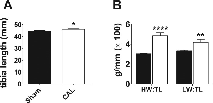
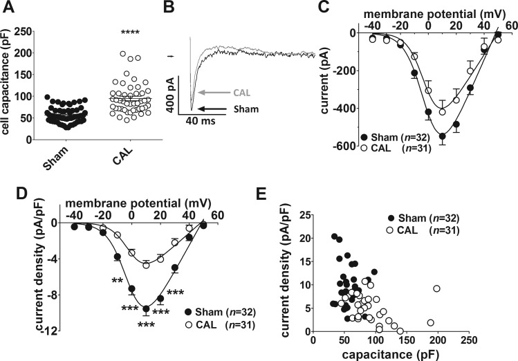
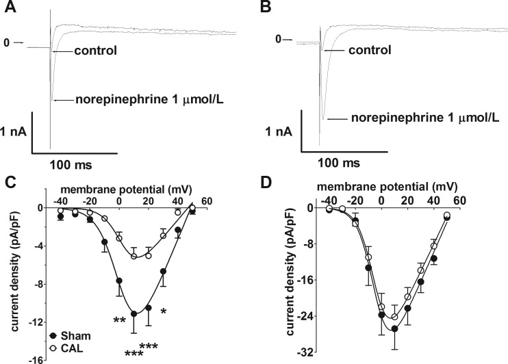
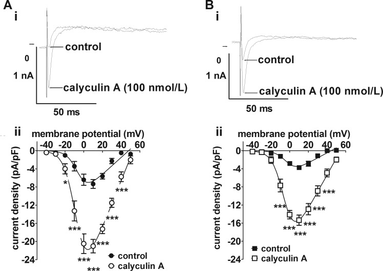
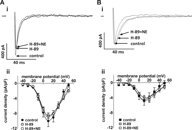
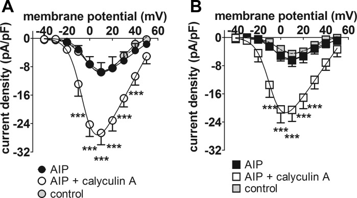
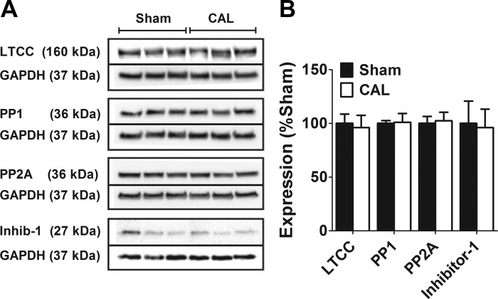
Constitutive regulation by PKA has recently been shown to contribute to L-type Ca2+ current (ICaL) at the ventricular t-tubule in heart failure. Conversely, reduction in constitutive regulation by PKA has been proposed to underlie the downregulation of atrial ICaL in heart failure. The hypothesis that downregulation of atrial ICaL in heart failure involves reduced channel phosphorylation was examined. Anesthetized adult male Wistar rats underwent surgical coronary artery ligation (CAL, N=10) or equivalent sham-operation (Sham, N=12). Left atrial myocytes were isolated ~18 wk postsurgery and whole cell currents recorded (holding potential=-80 mV). ICaL activated by depolarizing pulses to voltages from -40 to +50 mV were normalized to cell capacitance and current density-voltage relations plotted. CAL cell capacitances were ~1.67-fold greater than Sham (P ≤ 0.0001). Maximal ICaL conductance (Gmax ) was downregulated more than 2-fold in CAL vs. Sham myocytes (P < 0.0001). Norepinephrine (1 μmol/l) increased Gmax >50% more effectively in CAL than in Sham so that differences in ICaL density were abolished. Differences between CAL and Sham Gmax were not abolished by calyculin A (100 nmol/l), suggesting that increased protein dephosphorylation did not account for ICaL downregulation. Treatment with either H-89 (10 μmol/l) or AIP (5 μmol/l) had no effect on basal currents in Sham or CAL myocytes, indicating that, in contrast to ventricular myocytes, neither PKA nor CaMKII regulated basal ICaL Expression of the L-type α1C-subunit, protein phosphatases 1 and 2A, and inhibitor-1 proteins was unchanged. In conclusion, reduction in PKA-dependent regulation did not contribute to downregulation of atrial ICaL in heart failure.NEW & NOTEWORTHY Whole cell recording of L-type Ca2+ currents in atrial myocytes from rat hearts subjected to coronary artery ligation compared with those from sham-operated controls reveals marked reduction in current density in heart failure without change in channel subunit expression and associated with altered phosphorylation independent of protein kinase A.

Keywords: atrial remodeling; coronary artery ligation; voltage-gated Ca2+ channel.


Figures

Fig. 1.
Fig. 1.
Atrial L-type Ca2+ current density in heart failure. A: mean tibia length from 12 Sham and 10 CAL rats. *P < 0.05, Mann-Whitney test. B: mean heart weight/tibia length (HW/TL) and lung weight/tibia length (LW/TL) ratios. Sham, filled columns; CAL, open columns. **P < 0.01, ****P < 0.001, Mann-Whitney test.
Fig. 2.
Fig. 2.
A: whole cell capacitances for Sham (n = 63/12) and CAL (n = 47/10) myocytes. ****P < 0.0001, Mann-Whitney test. B: representative whole cell L-type Ca2+ current (ICaL) traces at +10 mV. Sham, black; CAL, gray. C: mean ICaL-voltage relations for Sham [filled circles, (n no. of cells/N no. of animals) = 32/12] and CAL (open circles, n/N = 31/10) myocytes. D: mean ICaL density-voltage relations for Sham (filled circles) and CAL (open circles) myocytes. Same data as shown in C. **P < 0.01, ***P < 0.001, 2-way repeated-measures ANOVA with Bonferroni posttest. Solid lines in C and D represent fits to Eq. 1. E: correlation between ICaL density at +10 mV in Sham (filled circles, n/N = 32/12) and CAL (open circles, n = 31/10) myocytes. Atrial ICaL density for the two groups of cells combined was significantly correlated (r = −0.4772, n/N = 63/22, P < 0.0001).
Fig. 3.
Fig. 3.
Effects of norepinephrine on atrial L-type Ca2+ current (ICaL) density. A: representative Ca2+ current traces at +10 mV in an atrial myocyte from a Sham-operated rat in control conditions and in the presence of 1 μmol/l norepinephrine. B: representative Ca2+ current traces in an atrial myocyte from a CAL rat in control conditions and in the presence of 1 μmol/l norepinephrine. Arrows indicate zero current level. C: mean ICaL density-voltage relations under control conditions from Sham-operated (n/N = 5/3, filled circles) and CAL (n/N = 9/4, open circles). *P < 0.05, **P < 0.01, ***P < 0.001, Bonferroni posttest following 2-way repeated-measures ANOVA. D: mean ICaL density-voltage relations in the presence of 1 μmol/l norepinephrine from Sham-operated (n/N = 5/3, open circles) and CAL (n/N = 9/4, filled circles). Data correspond to the control data shown in C. Note the difference in current density scale between C and D. Solid lines in C and D represent fits to Eq. 1.
Fig. 4.
Fig. 4.
Atrial L-type Ca2+ currents following phosphatase inhibition. A, i: representative current traces recorded at +10 mV from a Sham myocyte before and after superfusion with 100 nmol/l calyculin A. A, ii: ICaL density-voltage relations from Sham myocytes (n/N = 6/3) in control (filled circles) and in the presence of 100 nmol/l calyculin A (open circles). *P < 0.05, ***P < 0.001, 2-way repeated-measures ANOVA and Bonferroni posttest. B, i: representative current traces recorded at +10 mV from a CAL myocyte before and after superfusion with 100 nmol/l calyculin A. B, ii: ICaL density-voltage relations from CAL myocytes (n/N = 9/3) in control (filled squares) and in the presence of 100 nmol/l calyculin A (open squares). ***P < 0.001, 2-way ANOVA and Bonferroni posttest.
Fig. 5.
Fig. 5.
Effect of the protein kinase A inhibitor, H-89. A, i: representative Ca2+ current traces at +10 mV in an atrial myocyte from a Sham-operated rat in control Tyrode’s, in the presence of 10 μmol/l H-89, and in the presence of 10 μmol/l H-89 and 1 μmol/l norepinephrine (NE). A, ii: mean ICaL density-voltage relations from Sham myocytes (n/N = 8/4) in control (black-filled circles), in the presence of 10 μmol/l H-89 (gray-filled circles) and in the presence of 10 μmol/l H-89 and 1 μmol/l NE (open circles). B, i: representative Ca2+ current traces in an atrial myocyte from a CAL rat in control Tyrode’s, in the presence of 10 μmol/l H-89, and in the presence of 10 μmol/l H-89 and 1 μmol/l NE. Right-pointing arrows indicate zero current level. B, ii: mean ICaL density-voltage relations from CAL myocytes (n/N = 13/4) in control (black filled squares), in the presence of 10 μmol/l H-89 (gray-filled squares) and in the presence of 10 μmol/l H-89 and 1 μmol/l NE (open squares).
Fig. 6.
Fig. 6.
Effect of the Ca2+-calmodulin-dependent protein kinase II inhibitor, AIP. A: mean ICaL density-voltage relations for Sham myocytes with AIP included in the pipette solution (n/N = 4/1). Black-filled circles, AIP-dialyzed cells in control Tyrode’s; open circles, AIP-dialyzed cells in the presence of 100 nmol/l calyculin A. B: mean ICaL density-voltage relations for CAL myocytes with AIP included in the pipette solution (n/N = 5/1). Black-filled squares, AIP-dialyzed cells in control Tyrode’s; open squares, AIP-dialyzed cells in the presence of 100 nmol/l calyculin A. Data from Sham (A) and CAL (B) myocytes under control conditions (respectively, n/N = 33/12 and 31/10) are shown as, respectively, gray-filled circles and gray-filled squares, for comparison. ***P < 0.001, 2-way repeated-measures ANOVA and Bonferroni posttest vs. corresponding AIP control.
Fig. 7.
Fig. 7.
Left atrial expression of the Cav1.2 LTCC α1c subunit and various protein phosphatases in heart failure. A: representative Western blots of LTCC, protein phosphatases, and GAPDH from Sham and CAL rats. For each protein, the bands are taken from the same gel and have not been manipulated for contrast, color-balance, brightness, or background. Solid lines demarcate the bands for the target protein and for the GAPDH loading control, which were taken from the same gel. For each protein, the molecular weights of the bands are indicated. B: mean band intensity expressed relative to Sham as 100%. Data are means ± SE from 3 Sham and 3 CAL hearts.

References

    1. Aita Y, Kurebayashi N, Hirose S, Maturana AD. Protein kinase D regulates the human cardiac L-type voltage-gated calcium channel through serine 1884. FEBS Lett 585: 3903–3906, 2011. doi:10.1016/j.febslet.2011.11.011. - DOI - PubMed
    1. Andrade J, Khairy P, Dobrev D, Nattel S. The clinical profile and pathophysiology of atrial fibrillation: relationships among clinical features, epidemiology, and mechanisms. Circ Res 114: 1453–1468, 2014. doi:10.1161/CIRCRESAHA.114.303211. - DOI - PubMed
    1. Benjamin EJ, Chen P-S, Bild DE, Mascette AM, Albert CM, Alonso A, Calkins H, Connolly SJ, Curtis AB, Darbar D, Ellinor PT, Go AS, Goldschlager NF, Heckbert SR, Jalife J, Kerr CR, Levy D, Lloyd-Jones DM, Massie BM, Nattel S, Olgin JE, Packer DL, Po SS, Tsang TSM, Van Wagoner DR, Waldo AL, Wyse DG. Prevention of atrial fibrillation: report from a national heart, lung, and blood institute workshop. Circulation 119: 606–618, 2009. doi:10.1161/CIRCULATIONAHA.108.825380. - DOI - PMC - PubMed
    1. Boixel C, Gonzalez W, Louedec L, Hatem SN. Mechanisms of L-type Ca(2+) current downregulation in rat atrial myocytes during heart failure. Circ Res 89: 607–613, 2001. doi:10.1161/hh1901.096702. - DOI - PubMed
    1. Boixel C, Tessier S, Pansard Y, Lang-Lazdunski L, Mercadier J-J, Hatem SN. Tyrosine kinase and protein kinase C regulate L-type Ca(2+) current cooperatively in human atrial myocytes. Am J Physiol Heart Circ Physiol 278: H670–H676, 2000. - PubMed

MeSH terms

Substances

LinkOut - more resources