Comparative Study
. 2017 Jan;10(1):67-75.
doi: 10.1158/1940-6207.CAPR-16-0218. Epub 2016 Dec 6.

Whole-Exome Sequencing Validates a Preclinical Mouse Model for the Prevention and Treatment of Cutaneous Squamous Cell Carcinoma

Affiliations
Comparative Study

Whole-Exome Sequencing Validates a Preclinical Mouse Model for the Prevention and Treatment of Cutaneous Squamous Cell Carcinoma

Elena V Knatko et al. Cancer Prev Res (Phila). 2017 Jan.

Abstract

Cutaneous squamous cell carcinomas (cSCC) are among the most common and highly mutated human malignancies. Solar UV radiation is the major factor in the etiology of cSCC. Whole-exome sequencing of 18 microdissected tumor samples (cases) derived from SKH-1 hairless mice that had been chronically exposed to solar-simulated UV (SSUV) radiation showed a median point mutation (SNP) rate of 155 per Mb. The majority (78.6%) of the SNPs are C.G>T.A transitions, a characteristic UVR-induced mutational signature. Direct comparison with human cSCC cases showed high overlap in terms of both frequency and type of SNP mutations. Mutations in Trp53 were detected in 15 of 18 (83%) cases, with 20 of 21 SNP mutations located in the protein DNA-binding domain. Strikingly, multiple nonsynonymous SNP mutations in genes encoding Notch family members (Notch1-4) were present in 10 of 18 (55%) cases. The histopathologic spectrum of the mouse cSCC that develops in this model resembles very closely the spectrum of human cSCC. We conclude that the mouse SSUV cSCCs accurately represent the histopathologic and mutational spectra of the most prevalent tumor suppressors of human cSCC, validating the use of this preclinical model for the prevention and treatment of human cSCC. Cancer Prev Res; 10(1); 67-75. ©2016 AACR.


Conflict of interest statement

Disclosure of Potential Conflicts of Interest: None of the authors have any potential conflict of interest to disclose.

Figures

Figure 1
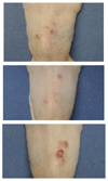
Figure 1. Macroscopic appearance of lesions that form in SKH-1 hairless mice following chronic exposure to solar-simulated UVR.
Female SKH-1 hairless mice were exposed chronically twice a week for 15 weeks to solar-simulated UVR (comprised of 2 J/cm2 UVA and 90 mJ/cm2 UVB) beginning at 8 weeks of age. The experiment was terminated 20 weeks after the end of the irradiation schedule. Examples of typical “field cancerization” of the skin of three animals at termination of the experiment (study week 35).
Figure 2
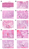
Figure 2. Histopathological spectrum of lesions that form in SKH-1 hairless mice following chronic exposure to solar-simulated UVR.
(A) Low grade (basal layer) epidermal dysplasia in perilesional skin; (B) Typical actinic keratosis with epidermal thickening associated with hyper and parakeratosis; (C) “Bowenoid” actinic keratosis amounting to carcinoma in situ; (D) higher power (400x) image of the same tumor (C) showing severe cytological atypia with loss of cell polarity, single cell dyskeratosis (arrow) and mitosis (arrow); (E) invasive SCC; note warty Bowenoid actinic keratosis overlying deeply invasive moderately differentiated carcinoma; (F) higher power (200x) image of the same tumor (E) showing islands of invasive carcinoma with stromal desmoplasia (arrow); (G) early invasion by SCC; (H) higher power (200x) image of the same tumor (I); note the irregular budding with a tongue of squamous cells extending into stroma (arrow), as well as single cell invasion (arrow); (I) invasive SCC abutting a band of skeletal muscle tissue; (J) higher power (200x) image of the same tumor (G) showing deep invasion by SCC cells encroaching onto skeletal muscle (arrows).
Figure 3
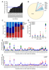
Figure 3. Mutational signature of solar-simulated UVR-induced mcSCC tumors.
(A) Total number of unique SNPs detected per tumor; the blue line indicates average across all tumors. (B) Pie chart of SNP frequencies indicating C>T (beige), C>G (purple), C>A (teal), C > T (green) A>T (green), A>G (blue), and A>C (white). (C) Frequency of base preceding each unique SNP mutation. (D) Mutation frequency of both transitions and transversions from hcSCC sequencing data from Pickering et al. (red circles) and South et al. (green circles), and in solar-simulated UVR-induced mcSCC tumors from this study (blue triangles). (E) Comparison of trinucleotide context of SNP mutations found in hcSCC reported by Pickering et al. (red dots), hcSCC reported by South et al. (green dots), and our mcSCC samples (blue triangles). (F) Comparison of trinucleotide context of SNP mutations found in a mouse UV-induced melanoma model (m-UV, red squares) and our mcSCC samples (blue triangles).
Figure 4
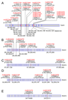
Figure 4. Schematic representation of Trp53 and Notch family nonsynonymous mutations in solar-simulated UVR-induced mcSCC tumors.
For all genes, exons are separated by blue lines starting with exon 1 on the left hand side. Mutations are indicated by red lines within the corresponding exons. Tumor case number, cDNA base pair change, and corresponding amino acid change are indicated above each mutation. Asterisks indicate a change to a stop codon. Numbers below each mutation indicate chromosome location. Location of unique non synonymous mutations in Trp53 (A), Notch1 (B), Notch2 (C), Notch3 (D), and Notch4 (E).

References

    1. Alam M, Ratner D. Cutaneous squamous-cell carcinoma. N Engl J Med. 2001;344:975–83. - PubMed
    1. Jennings L, Schmults CD. Management of high-risk cutaneous squamous cell carcinoma. J Clin Aesthet Dermatol. 2010;3:39–48. - PMC - PubMed
    1. (www.isdscotland.org/Health-Topics/Cancer/Cancer-Statistics/Skin/#squamousDavid)

    1. Karia PS, Han J, Schmults CD. Cutaneous squamous cell carcinoma: estimated incidence of disease, nodal metastasis, and deaths from disease in the United States, 2012. J Am Acad Dermatol. 2013;68:957–66. - PubMed
    1. Levine DE, Karia PS, Schmults CD. Outcomes of Patients With Multiple Cutaneous Squamous Cell Carcinomas: A 10-Year Single-Institution Cohort Study. JAMA Dermatology. 2015;151:1220–5. - PubMed

MeSH terms

Substances