

β-Lapachone Inhibits Lung Metastasis of Colorectal Cancer by Inducing Apoptosis of CT26 Cells
- PMID: 27923905
- PMCID: PMC5739146
- DOI: 10.1177/1534735416681638
β-Lapachone Inhibits Lung Metastasis of Colorectal Cancer by Inducing Apoptosis of CT26 Cells
Abstract
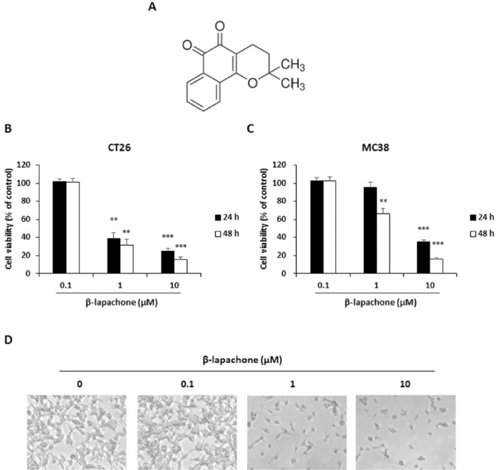
Background: β-Lapachone is a quinone-containing compound found in red lapacho ( Tabebuia impetiginosa, syn. T avellanedae) trees. Lapacho has been used in traditional medicine by several South and Central American indigenous people to treat various types of cancer. The purpose of this study was to investigate the antimetastatic properties of β-lapachone and the underlying mechanisms using colon cancer cells.
Methods: This research used metastatic murine colon cancer cell lines, colon 26 (CT26) and colon 38 (MC38). A WST assay, annexin V assay, cell cycle analysis, wound healing assay, invasion assay, western blot analysis, and real-time reverse transcription-polymerase chain reaction were performed to examine the effects of β-lapachone on metastatic phenotypes and molecular mechanisms. The effect of β-lapachone on lung metastasis was assessed in a mouse experimental metastasis model.
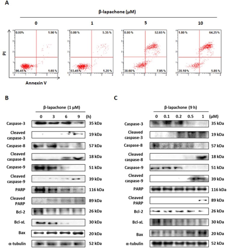
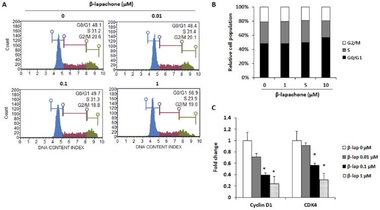
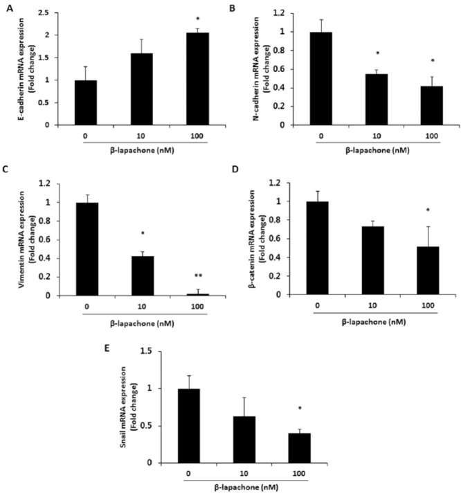
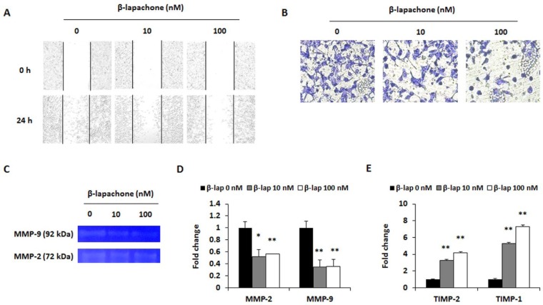
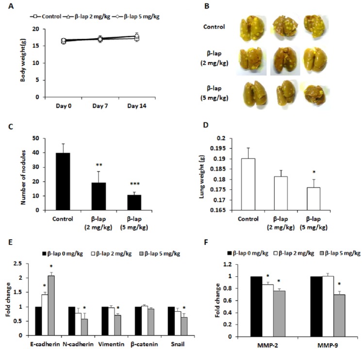
Results: We found that the inhibition of proliferation of the colon cancer cell lines by β-lapachone was due to the induction of apoptosis and cell cycle arrest. β-Lapachone induced the apoptosis of CT26 cells through caspase-3, -8, and -9 activation; poly(ADP-ribose) polymerase cleavage; and downregulation of the Bcl-2 family in a dose- and time-dependent manner. In addition, a low concentration of β-lapachone decreased the cell migration and invasion by decreasing the expression of matrix metalloproteinases-2 and -9, and increased the expression of tissue inhibitors of metalloproteinases-1 and -2. Moreover, β-lapachone treatment regulated the expression of epithelial-mesenchymal transition markers such as E- and N-cadherin, vimentin, β-catenin, and Snail in CT26 cells. In the mouse experimental metastasis model, β-lapachone significantly inhibited the lung metastasis of CT26 cells.
Conclusions: Our results demonstrated the inhibitory effect of β-lapachone on colorectal lung metastasis. This compound may be useful for developing therapeutic agents to treat metastatic cancer.
Keywords: CT26 cells; apoptosis; colorectal cancer; lung metastasis; β-lapachone.
Conflict of interest statement
Figures
Similar articles
-
Inhibitory effect of quercetin on colorectal lung metastasis through inducing apoptosis, and suppression of metastatic ability.Phytomedicine. 2016 Dec 1;23(13):1680-1690. doi: 10.1016/j.phymed.2016.09.011. Epub 2016 Oct 1. Phytomedicine. 2016. PMID: 27823633
-
Beta-lapachone, a quinone isolated from Tabebuia avellanedae, induces apoptosis in HepG2 hepatoma cell line through induction of Bax and activation of caspase.J Med Food. 2006 Summer;9(2):161-8. doi: 10.1089/jmf.2006.9.161. J Med Food. 2006. PMID: 16822200
-
beta-lapachone induces growth inhibition and apoptosis in bladder cancer cells by modulation of Bcl-2 family and activation of caspases.Exp Oncol. 2006 Mar;28(1):30-5. Exp Oncol. 2006. PMID: 16614704
-
[Cytotoxicity of beta-lapachone, an naphthoquinone with possible therapeutic use].Medicina (B Aires). 2001;61(3):343-50. Medicina (B Aires). 2001. PMID: 11474885 Review. Spanish.
-
Therapeutic potential of ethoxy mansonone G: A comprehensive exploration of its anticancer actions in breast cancer, colorectal cancer, and non-small cell lung carcinoma.Cell Biol Int. 2024 Sep;48(9):1229-1239. doi: 10.1002/cbin.12207. Epub 2024 Jun 23. Cell Biol Int. 2024. PMID: 38924324 Review.
Cited by
-
Metabolic profiling of cytotoxic metabolites from five Tabebuia species supported by molecular correlation analysis.Sci Rep. 2021 Apr 16;11(1):8405. doi: 10.1038/s41598-021-87695-w. Sci Rep. 2021. PMID: 33863934 Free PMC article.
-
NQO1 is Required for β-Lapachone-Mediated Downregulation of Breast-Cancer Stem-Cell Activity.Int J Mol Sci. 2018 Nov 30;19(12):3813. doi: 10.3390/ijms19123813. Int J Mol Sci. 2018. PMID: 30513573 Free PMC article.
-
X-ray repair cross-complementing protein 1 (XRCC1) loss promotes β-lapachone -induced apoptosis in pancreatic cancer cells.BMC Cancer. 2021 Nov 17;21(1):1234. doi: 10.1186/s12885-021-08979-y. BMC Cancer. 2021. PMID: 34789190 Free PMC article.
-
In Vitro Antiproliferative Activity in Plants of the Genus Tabebuia: A Systematic Review.Molecules. 2025 May 25;30(11):2315. doi: 10.3390/molecules30112315. Molecules. 2025. PMID: 40509203 Free PMC article. Review.
-
Liposomal β-Sitosterol Suppresses Metastasis of CT26/luc Colon Carcinoma via Inhibition of MMP-9 and Evoke of Immune System.Pharmaceutics. 2022 Jun 7;14(6):1214. doi: 10.3390/pharmaceutics14061214. Pharmaceutics. 2022. PMID: 35745788 Free PMC article.
References
-
- Jemal A, Bray F, Center MM, Ferlay J, Ward E, Forman D. Global cancer statistics. CA Cancer J Clin. 2011;61:69-90. - PubMed
-
- Seymour MT, Stenning SP, Cassidy J. Attitudes and practice in the management of metastatic colorectal cancer in Britain. Colorectal Cancer Working Party of the UK Medical Research Council. Clin Oncol (R Coll Radiol). 1997;9:248-251. - PubMed
-
- Labianca R, Beretta GD, Kildani B, et al. Colon cancer. Crit Rev Oncol Hematol. 2010;74:106-133. - PubMed
MeSH terms
Substances
LinkOut - more resources
Full Text Sources
Other Literature Sources
Medical
Research Materials
Miscellaneous
