. 2017 Jan;214(1):17-25.
doi: 10.1084/jem.20160447. Epub 2016 Dec 6.

Estrogen activation of microglia underlies the sexually dimorphic differences in Nf1 optic glioma-induced retinal pathology

Affiliations

Estrogen activation of microglia underlies the sexually dimorphic differences in Nf1 optic glioma-induced retinal pathology

Joseph A Toonen et al. J Exp Med. 2017 Jan.

Abstract

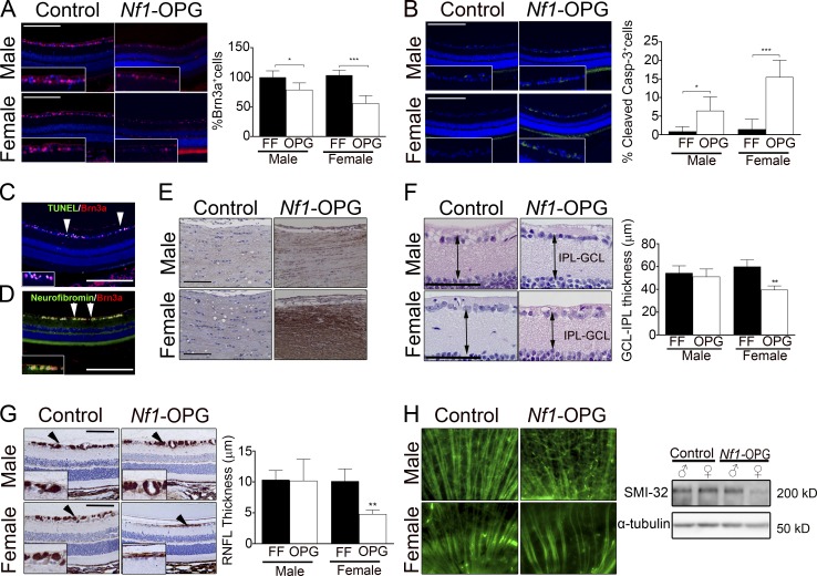
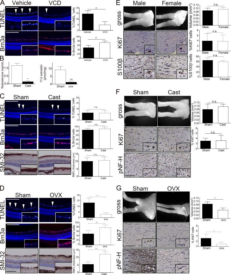
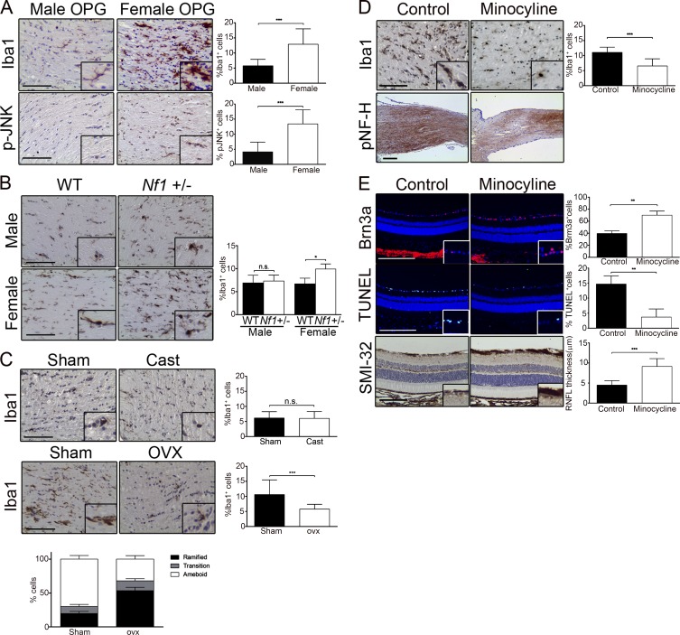
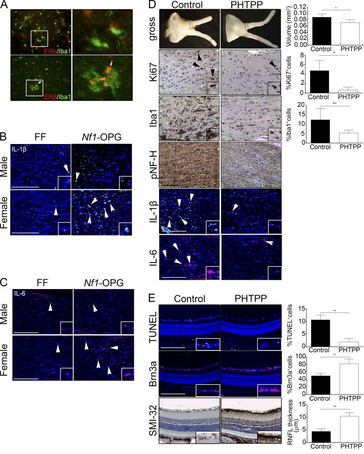
Children with neurofibromatosis type 1 (NF1) develop low-grade brain tumors throughout the optic pathway. Nearly 50% of children with optic pathway gliomas (OPGs) experience visual impairment, and few regain their vision after chemotherapy. Recent studies have revealed that girls with optic nerve gliomas are five times more likely to lose vision and require treatment than boys. To determine the mechanism underlying this sexually dimorphic difference in clinical outcome, we leveraged Nf1 optic glioma (Nf1-OPG) mice. We demonstrate that female Nf1-OPG mice exhibit greater retinal ganglion cell (RGC) loss and only females have retinal nerve fiber layer (RNFL) thinning, despite mice of both sexes harboring tumors of identical volumes and proliferation. Female gonadal sex hormones are responsible for this sexual dimorphism, as ovariectomy, but not castration, of Nf1-OPG mice normalizes RGC survival and RNFL thickness. In addition, female Nf1-OPG mice have threefold more microglia than their male counterparts, and minocycline inhibition of microglia corrects the retinal pathology. Moreover, pharmacologic inhibition of microglial estrogen receptor-β (ERβ) function corrects the retinal abnormalities in female Nf1-OPG mice. Collectively, these studies establish that female gonadal sex hormones underlie the sexual dimorphic differences in Nf1 optic glioma-induced retinal dysfunction by operating at the level of tumor-associated microglial activation.


Figures

Figure 1.
Figure 1.
Retinal dysfunction is sexually dimorphic in Nf1-OPG mice. (A) The percentage of Brn3a+ RGCs was decreased in female Nf1-OPG mice (mean, 56.13 ± 13.11% SD; n = 7 mice) compared with FF controls (103.6 ± 8.6%; n = 7 mice), and to a lesser degree in their male counterparts (FF males, 100 ± 10.91%; n = 7 mice; Nf1-OPG males, 79.01 ± 11.6%; n = 7 mice). (B) Cleaved caspase-3+ cells were increased in female Nf1-OPG mice (15.52 ± 4.52%; n = 6 mice) compared with FF controls (1.45 ± 1.09%; n = 6 mice), but were less elevated in male Nf1-OPG mice (0.822 ± 1.29%; n = 8 mice) relative to FF controls (6.451 ± 3.72%; n = 6 mice). (C) Double-labeled Brn3a+ (red) and TUNEL+ cells (green) were detected in female Nf1-OPG mice. (D) Neurofibromin (green) was primarily expressed in Brn3a+ (red) RGCs in WT mice. (E) pNF-H immunostaining was detected in the optic nerves of female Nf1-OPG mice, but not Nf1-OPG males or controls. (F) IPL-GCL layer thickness showed no differences in males (FF, 54.41 ± 6.3 μm; n = 6 mice; OPG, 51.23 ± 6.67 μm; n = 6 mice), whereas thinning was found in Nf1-OPG females (39.54 ± 3.55 μm; n = 7 mice) compared with controls (FF, 60.1 ± 5.96 μm; n = 6 mice). (G) SMI-32 staining (RNFL thickness) revealed thinning in female Nf1-OPG retinae (4.82 ± 0.64 μm; n = 6 mice) relative to controls (10.14 ± 1.99 μm; n = 6 mice). No differences were seen in males (FF, 10.37 ± 1.5 μm; n = 6 mice; OPG, 10.22 ± 3.46 μm; n = 6 mice). (H) Decreased SMI-32 staining was observed in female Nf1-OPG retinal flatmounts (left) and by immunoblotting (right). Data in A, B, F, and G were analyzed using a one-way ANOVA (Kruskal-Wallis test) with a Dunn’s multiple comparison post-test. Similar results were obtained in a second independent experiment containing six mice per group. Bars, 100 µm. *, P < 0.05; **, P < 0.01; ***, P < 0.001.
Figure 2.
Figure 2.
Depletion of female gonadal sex hormones rescues tumor growth and RGC death in Nf1-OPG females. (A) VCD treatment decreased TUNEL+ cells in the RGC layer (Vehicle, 14.8 ± 2.7%; VCD, 4.9 ± 2.9; n = 6 mice) and increased the percentage of Brn3a+ RGCs in FMC mice (Vehicle, 49.3 ± 10.8%; VCD, 73.1 ± 7.5; n = 7 mice/group). (B) Serum testosterone levels decreased after castration (Cast) of male Nf1-OPG mice (Sham, 1.19 ±0.14 ng/ml; Cast, 0.14 ± 0.05 ng/ml; n = 5 mice/group). Serum 17-β estradiol levels decreased after OVX of female Nf1-OPG mice (Sham, 9.33 ± 2.1 pmol/ml; OVX, 1.29 ± 0.88 pmol/ml; n = 7 mice/group). (C) Castration did not change retinal apoptosis (%TUNEL+ cells; Sham, 6.53 ± 1.83; Cast, 6.25 ± 2.16; n = 7 mice), RGC loss (%Brn3a+ cells; Sham, 83.78 ± 6.7; Cast, 86.21 ± 8.1; n = 7 mice/group), or RNFL thickness (SMI-32 staining; Sham, 10.14 ± 2.35 μm; Cast, 10.77 ± 2.2 μm; n = 7 mice/group). (D) OVX reduced RGC death (%TUNEL+ cells; Sham, 13.19 ± 4.0; OVX, 2.98 ± 1.2; n = 7 mice/group), RGC loss (%Brn3a; Sham, 49.56 ± 13.9; OVX, 77.42 ± 11.7; n = 7 mice/group), and RNFL thinning (SMI-32 staining; Sham, 5.51 ± 1.6 μm; OVX, 12.2 ± 1.97 μm; n = 7 mice/group). (E) No differences in optic nerve volumes (Male, 0.087 ± 0.003 mm3; Female, 0.095 ± 0.011 mm3; n = 17 mice/group), %Ki67+ cells (Male, 3.95 ± 2.81%; Female 3.76 ± 0.81%; n = 9 mice/group), or percentage of S100β+ cells (Male, 65.35 ± 5.01%; Female, 59.81 ± 5.1%; n = 5 mice/group) were found in the optic nerves of male compared with female Nf1-OPG mice. (F) Optic nerve volume measurements (top; Sham, 0.084 ± 0.02 mm3; Cast, 0.092 ± 0.02 mm3; n = 6 mice/group), %Ki67+ cells (middle; Sham, 3.08 ± 1.1%; Cast, 3.19 ± 2.08%; n = 6 mice/group), and pNF-H staining (bottom) within the optic nerve were unchanged after castration. (G) Optic nerve volumes (top; Sham, 0.085 ± 0.006 mm3; OVX, 0.049 ± 0.004 mm3; n = 5 mice/group) and %Ki67+ cells (middle; Sham, 3.2 ± 0.64%; OVX, 0.96 ± 0.34%; n = 5 mice/group) were decreased after OVX. Axonal damage (pNF-H, bottom) was decreased after OVX of Nf1-OPG females relative to sham controls. Data in A–G were analyzed using a nonparametric Student’s t test (Mann-Whitney). Similar results were obtained in a second independent experiment containing six mice per group. Bars, 100 µm. *, P < 0.05; **, P < 0.01; ***, P < 0.001; n.s., not significant.
Figure 3.
Figure 3.
Microglia causes axonal damage and RGC death in female Nf1-OPG mice. (A) Optic nerves of female Nf1-OPG mice contained increased %Iba1+ cells (Male, 5.8 ± 2.11%; Female, 12.96 ± 5.06%; n = 10 mice/group) and p-JNK+ cells (Male, 4.16 ± 3.2%; Female, 13.5 ± 4.7%; n = 8 mice/group). (B) Immunostaining revealed increased Iba1+ cells in Nf1+/− females compared with WT controls (WT, 6.7 ± 1.1%; Nf1+/−, 9.9 ± 1.1%; n = 6 mice/group); however, no differences were observed in males (WT, 6.9 ± 1.7%; Nf1+/−, 7.4 ± 1.3%). (C) The %Iba1+ cells (top; Sham, 6.21 ± 2.05%; Cast, 6.1 ± 2.26%; n = 8 mice/group) did not differ between castrated Nf1-OPG males and sham controls. In contrast, the %Iba1+ cells (bottom; Sham, 10.7 ± 4.84%; OVX, 5.83 ± 1.55%; n = 11 mice/group) were decreased after OVX in Nf1-OPG females compared with sham controls. Ramified microglia constitute 19.8% of the total microglia in sham Nf1-OPG female mice and 53.6% of the total microglia after OVX, whereas amoeboid or activated microglia are constituted 70% of the total microglia in sham Nf1-OPG optic nerves and 32% in OVX optic nerves (n = 6 mice/group). (D) Minocycline-treated female Nf1-OPG mice exhibited a decreased %Iba1+ cells in the optic nerve (Control, 11.12 ± 1.71%; minocycline, 6.61 ± 2.3%; n = 8 mice/group) and axonal damage (pNF-H immunostaining). (E) After minocycline treatment, there was a reduction in the %TUNEL+ cells (Control, 14.75 ± 1.1; minocycline, 3.75 ± 1.82; n = 6 mice/group), an increase in the %Brn3a+ cells (Control, 39.79 ± 1.69%; minocycline, 70.42 ± 2.66%; n = 6 mice/group), and increased the RNFL thickness (SMI-32 staining; Control, 4.51 ± 1.1 μm; minocycline, 9.2 ± 1.9 μm; n = 8 mice/group). Data in A–E were analyzed using a nonparametric Student’s t test (Mann-Whitney). Similar results were obtained in a second independent experiment containing five mice per group. Bars, 100 µm. *, P < 0.05; **, P < 0.01; ***, P < 0.001. n.s., not significant.
Figure 4.
Figure 4.
Estrogen acts through ERβ to activate optic nerve microglia in female Nf1-OPG mice. (A) Immunostaining for Iba1+, ERα+ double-labeled cells in the optic nerve (bottom, arrows). Immunostaining was performed for Il-1β (B) and Il-6 (C) in the optic nerves of FF male and female mice, as well as in Nf1-OPG male and female mice. (D) Optic nerves volumes (Control, 0.089 ± 0.02 mm3; PHTPP, 0.071 ± 0.01 mm3; n = 7 mice/group) and %Ki67+ cells (Control, 4.75 ± 2.2%; PHTPP, 0.64 ± 0.59%; n = 6 mice/group) were decreased after PHTPP treatment (top). Microglia numbers (%Iba1+ cells, Control, 12.33 ± 5.9; PHTPP, 5.44 ± 1.3, n = 8 mice/group), axonal injury (pNF-H), and IL-1β/IL-6 levels were decreased after treatment with PHTPP. (E) PHTPP treatment reduced the %TUNEL+ cells (Control, 10.7 ± 0.78%; PHTPP, 1.95 ± 0.56%; n = 6 mice/group), increased the %Brn3a+ cells (Control, 49.58 ± 2.69%; PHTPP, 81.7 ± 4.9%; n = 6 mice/group), and increased the RNFL thickness (SMI-32 staining; Control, 4.5 ± 0.4 μm; PHTPP, 10.5 ± 0.5 μm; n = 6 mice/group). Data in D and E were analyzed using a nonparametric Student’s t test (Mann-Whitney). Similar results were obtained in a second independent experiment containing five mice per group. Bars, 100 µm. *, P < 0.05; **, P < 0.01; ***, P < 0.001. n.s., not significant.

Similar articles

Cited by

References

    1. Arnold A.P. 2014. Conceptual frameworks and mouse models for studying sex differences in physiology and disease: why compensation changes the game. Exp. Neurol. 259:2–9. 10.1016/j.expneurol.2014.01.021 - DOI - PMC - PubMed
    1. Avery R.A., Liu G.T., Fisher M.J., Quinn G.E., Belasco J.B., Phillips P.C., Maguire M.G., and Balcer L.J.. 2011. Retinal nerve fiber layer thickness in children with optic pathway gliomas. Am. J. Ophthalmol. 151:542–9.e2. 10.1016/j.ajo.2010.08.046 - DOI - PMC - PubMed
    1. Azevedo E.P., Ledo J.H., Barbosa G., Sobrinho M., Diniz L., Fonseca A.C., Gomes F., Romão L., Lima F.R., Palhano F.L., et al. . 2013. Activated microglia mediate synapse loss and short-term memory deficits in a mouse model of transthyretin-related oculoleptomeningeal amyloidosis. Cell Death Dis. 4:e789 10.1038/cddis.2013.325 - DOI - PMC - PubMed
    1. Bajenaru M.L., Hernandez M.R., Perry A., Zhu Y., Parada L.F., Garbow J.R., and Gutmann D.H.. 2003. Optic nerve glioma in mice requires astrocyte Nf1 gene inactivation and Nf1 brain heterozygosity. Cancer Res. 63:8573–8577. - PubMed
    1. Bondesson M., Hao R., Lin C.Y., Williams C., and Gustafsson J.A.. 2015. Estrogen receptor signaling during vertebrate development. Biochim. Biophys. Acta. 1849:142–151. 10.1016/j.bbagrm.2014.06.005 - DOI - PMC - PubMed

Publication types