. 2016 Dec 6;9(457):ra119.
doi: 10.1126/scisignal.aaj1460.

Widespread control of calcium signaling by a family of SERCA-inhibiting micropeptides

Affiliations

Widespread control of calcium signaling by a family of SERCA-inhibiting micropeptides

Douglas M Anderson et al. Sci Signal. .

Abstract

Micropeptides function as master regulators of calcium-dependent signaling in muscle. Sarco/endoplasmic reticulum Ca2+ ATPase (SERCA), the membrane pump that promotes muscle relaxation by taking up Ca2+ into the sarcoplasmic reticulum, is directly inhibited by three muscle-specific micropeptides: myoregulin (MLN), phospholamban (PLN), and sarcolipin (SLN). The widespread and essential function of SERCA across diverse cell types has raised questions as to how SERCA is regulated in cells that lack MLN, PLN, and SLN. We identified two transmembrane micropeptides, endoregulin (ELN) and another-regulin (ALN), that share key amino acids with their muscle-specific counterparts and function as direct inhibitors of SERCA pump activity. The distribution of transcripts encoding ELN and ALN mirrored that of SERCA isoform-encoding transcripts in nonmuscle cell types. Our findings identify additional members of the SERCA-inhibitory micropeptide family, revealing a conserved mechanism for the control of intracellular Ca2+ dynamics in both muscle and nonmuscle cell types.


Conflict of interest statement

Competing interests: The authors declare that they have no competing interests.

Figures

Fig. 1
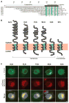
Fig. 1. Discovery of transmembrane micropeptides related to MLN, PLN, and SLN
(A) Sequence alignment of the vertebrate micropeptides ALN, ELN, PLN, MLN, and SLN from mouse and SCL from D. melanogaster highlights the residues that they share in common in their transmembrane helices. “*” denotes identically conserved residues, and “.” denotes weakly similar residues. (B) ALN and ELN encode single C-terminal transmembrane α helices and are predicted to insert into intracellular membranes similar to MLN, PLN, and SLN. The residues that they share in common, which mediate SERCA binding, are arranged along a single helical face. (C) Colocalization of FLAG-tagged micropeptides and mCherry-SERCA2b in the ER of COS-7 cells. Images are representative of two independent experiments. Scale bar, 5 μm.
Fig. 2
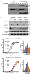
Fig. 2. ALN and ELN bind directly to and inhibit SERCA pump activity
(A and B) Coimmunoprecipitation experiments with HA-tagged micropeptides and a Myc-tagged SERCA2b using transient cotransfections in COS-7 cells in the absence (A) or presence (B) of competing GFP-tagged PLN. Immunoblots are representative of three independent experiments. (C and D) Oxalate-supported Ca2+-dependent Ca2+ uptake assays demonstrating the inhibitory function of micropeptide expression on SERCA2b (C) and SERCA3a (D). Vmax and KCa values were calculated from 12 independent measurements for each sample using an unpaired t test. Bar graphs depict the calculated average KCa ± SEM (six independent experiments). *P < 0.05; **P < 0.01.
Fig. 3
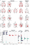
Fig. 3. Tissue and cell-type specific expression of micropeptides and SERCA isoforms in the developing and adult mouse
(A and B) In situ hybridization of antisense RNA probes specific for major SERCA iso-forms and micropeptides during mouse embryonic day E14.5 transverse (A) and E15.5 midsagittal (B). skm, skeletal muscle; at, atria; vt, ventricle; lu, lung; br, bronchus; to, tongue; dia, diaphragm; sg, salivary gland; bf, brown fat; liv, liver; pa, pancreas; in, intestine; bd, bladder; ep, epidermal epithelium. Scale bar, 1 mm. Images are representative of two independent experiments. (C) Quantitative reverse transcription polymerase chain reaction (qRT-PCR) showing relative expression of major SERCA isoforms and micropeptides across adult mouse tissues. Quad, quadriceps; TA, tibi-alis anterior; EDL, extensor digitorum longus; FB, fibroblasts; MB, myoblasts; C2C12 +1, +2, +3, C2C12 differentiation day 1, day 2, and day 3. Bar graphs depict means ± SEM from a single experiment (representative of three independent experiments).
Fig. 4
Fig. 4. A family of SERCA-inhibiting micropeptides
Model depicting the expression patterns of the predominant SERCA and micropeptide inhibitors across different muscle and nonmuscle tissues in vertebrates. The discovery of ELN and ALN suggests that the regulation of SERCA activity by SERCA-inhibiting micropeptides represents a general mechanism to control calcium handling across diverse cell types.

Similar articles

Cited by

References

    1. Rizzuto R, Pozzan T. Microdomains of intracellular Ca2+: Molecular determinants and functional consequences. Physiol Rev. 2006;86:369–408. - PubMed
    1. Berridge MJ. Unlocking the secrets of cell signaling. Annu Rev Physiol. 2005;67:1–21. - PubMed
    1. Clapham DE. Calcium signaling. Cell. 2007;131:1047–1058. - PubMed
    1. Berridge MJ, Bootman MD, Roderick HL. Calcium signalling: Dynamics, homeostasis and remodelling. Nat Rev Mol Cell Biol. 2003;4:517–529. - PubMed
    1. Rossi AE, Dirksen RT. Sarcoplasmic reticulum: The dynamic calcium governor of muscle. Muscle Nerve. 2006;33:715–731. - PubMed

MeSH terms