. 2017 Apr 20;45(7):4068-4080.
doi: 10.1093/nar/gkw1225.

Regulation of Prp43-mediated disassembly of spliceosomes by its cofactors Ntr1 and Ntr2

Affiliations

Regulation of Prp43-mediated disassembly of spliceosomes by its cofactors Ntr1 and Ntr2

Jean-Baptiste Fourmann et al. Nucleic Acids Res. .

Abstract

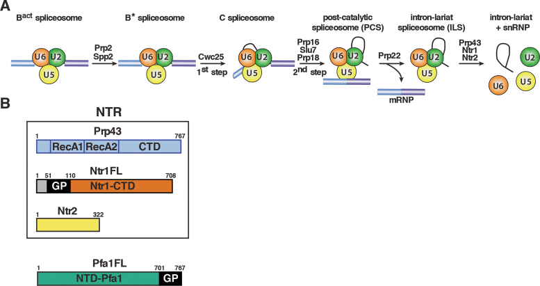
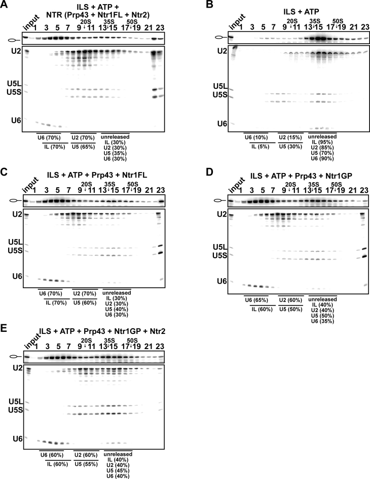
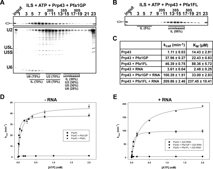
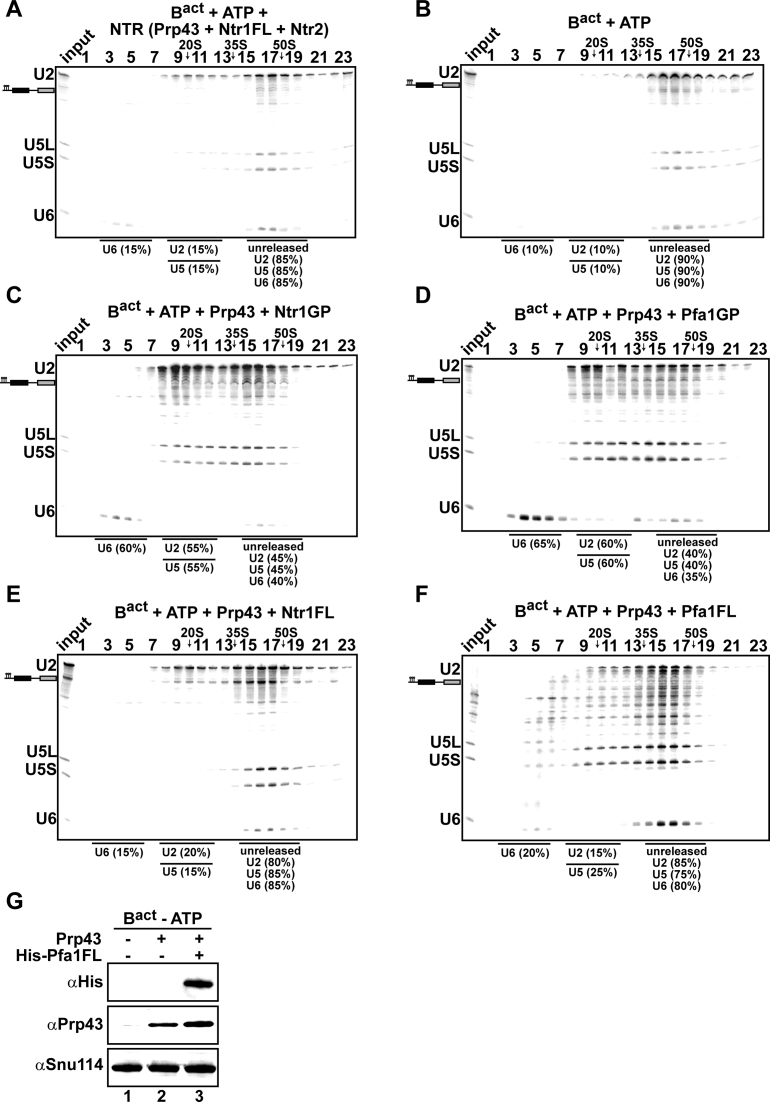
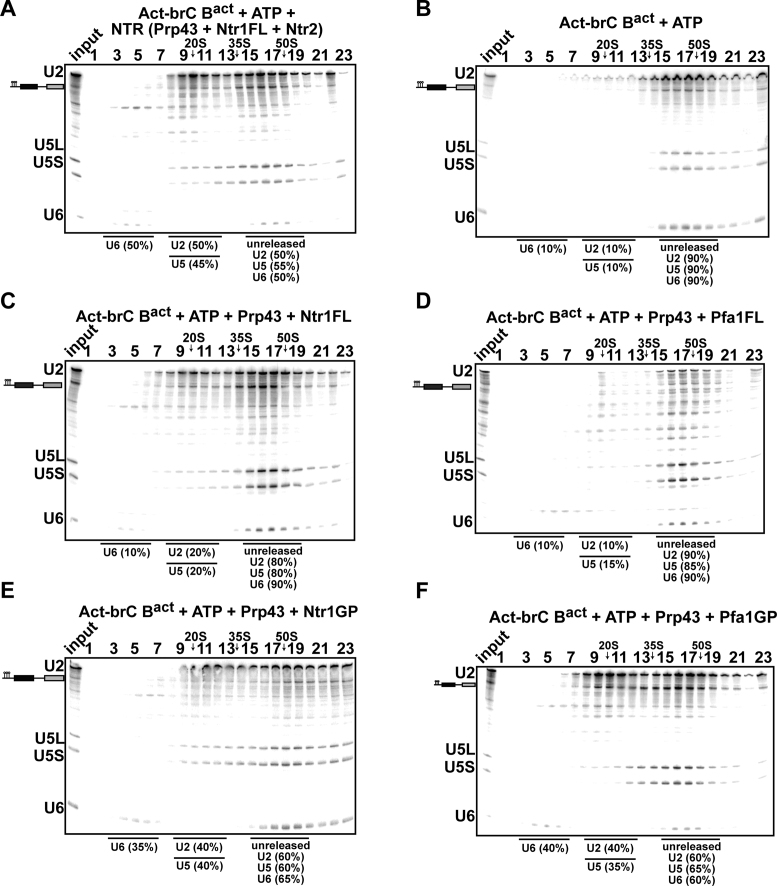
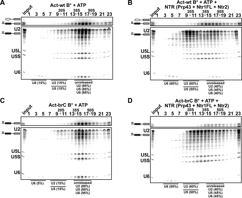
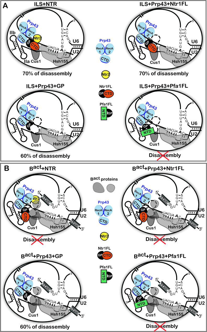
The DEAH-box NTPase Prp43 disassembles spliceosomes in co-operation with the cofactors Ntr1/Spp382 and Ntr2, forming the NTR complex. How Prp43 is regulated by its cofactors to discard selectively only intron-lariat spliceosomes (ILS) and defective spliceosomes and to prevent disassembly of earlier and properly assembled/wild-type spliceosomes remains unclear. First, we show that Ntr1΄s G-patch motif (Ntr1GP) can be replaced by the GP motif of Pfa1/Sqs1, a Prp43΄s cofactor in ribosome biogenesis, demonstrating that the specific function of Ntr1GP is to activate Prp43 for spliceosome disassembly and not to guide Prp43 to its binding site in the spliceosome. Furthermore, we show that Ntr1΄s C-terminal domain (CTD) plays a safeguarding role by preventing Prp43 from disrupting wild-type spliceosomes other than the ILS. Ntr1 and Ntr2 can also discriminate between wild-type and defective spliceosomes. In both type of spliceosomes, Ntr1-CTD impedes Prp43-mediated disassembly while the Ntr1GP promotes disassembly. Intriguingly, Ntr2 plays a specific role in defective spliceosomes, likely by stabilizing Ntr1 and allowing Prp43 to enter a productive interaction with the GP motif of Ntr1. Our data indicate that Ntr1 and Ntr2 act as 'doorkeepers' and suggest that both cofactors inspect the RNP structure of spliceosomal complexes thereby targeting suboptimal spliceosomes for Prp43-mediated disassembly.


Figures

Figure 1.
Figure 1.
Isolation of intron-lariat spliceosomes (ILSs) and schematic representation of the NTR complex and the G-patch cofactor Pfa1. (A) Activated spliceosomes (BactΔPrp2) assembled on Actin7 wild-type pre-mRNA in heat-inactivated splicing extracts from a prp2-1 yeast strain expressing a temperature-sensitive Prp2 mutant, were first purified. Purified BactΔPrp2 complexes were then incubated with recombinant Prp2 and Spp2, generating the B* spliceosome, and then Cwc25 was added to promote catalysis of step 1 of splicing and the formation of complex C. For catalysis of step 2, which generates post-catalytic spliceosomes (PCS), recombinant Prp16, Slu7 and Prp18 were added. Finally, for the purification of the ILS, the spliced mRNA was dissociated from the ILS by incubation of the PCS with Prp22 and ATP. Addition of ATP and recombinant Prp43, Ntr1 and Ntr2, leads to disassembly of the ILS into the intron-lariat, 20S U2 snRNP, 18S U5 snRNP and free U6 snRNA (26). (B) The NTR complex is composed of Prp43, Ntr1 and Ntr2. Full-length Ntr1 (Ntr1FL), includes the G-patch motif (GP) at its N-terminal region (residues 51–110), and the C-terminal domain (Ntr1-CTD), Pfa1 full-length (Pfa1FL) includes the G-patch motif (GP) at its C-terminal region (residues 701 to 767), and a N-terminal domain (NTD-Pfa1).
Figure 2.
Figure 2.
Full-length Ntr1 is sufficient to promote dissociation of the ILS by Prp43. 10–30% glycerol gradient sedimentation of purified ILS incubated in solution with (A) ATP plus NTR, (B) no recombinant protein, (C) Prp43 and full-length Ntr1 (Ntr1FL), (D) Prp43 and Ntr1 G-patch motif (Ntr1GP), (E) Prp43, Ntr1GP and Ntr2. U2, U5 and U6 snRNAs were visualized by northern blotting followed by autoradiography. RNA identities are indicated on the left. Quantifications were performed with ImageQuant software (Molecular Dynamics). ILS: numbers represent the percentage of intron-lariat RNA released in the peak fractions (sum of fractions 3–7) or associated with the ILS (unreleased, sum of fractions 13–17) relative to the intron-lariat RNA distributed in fractions 3–7 and 13–17, the sum of which was set to 100%. U2 and U5: numbers represent the percentage of U2 or U5 snRNAs released in the peak fractions (sum of fractions 8–12) or associated with the ILS (unreleased, sum of fractions 13–17) relative to the U2 or U5 snRNA distributed in the fractions 8–12 and 13–17, the sum of which was set to 100%. U6: numbers represent the percentage of U6 snRNA released in the peak fractions (sum of fractions 2–6) or associated with the ILS (unreleased, sum of fractions 13–17) relative to the U6 snRNA distributed in the fractions 2–6 and 13–17, the sum of which was set to 100%.
Figure 3.
Figure 3.
Prp43 dissociates the ILS in co-operation with the GP motif of Pfa1 but not with full-length Pfa1. 10–30% glycerol gradient sedimentation of purified ILS incubated in solution with ATP, Prp43 and (A) Pfa1 G-patch motif (Pfa1GP), (B) Pfa1 full length (Pfa1FL). Samples were analyzed and quantified as described in Figure 2. (C) Kinetic parameters (kcat and KM) of Prp43, Prp43 + Pfa1GP and Prp43 + Pfa1FL in absence or presence of poly-A20 RNA cofactor. (D) ATPase activity of Prp43 in the absence and presence of Pfa1GP and Pfa1FL, respectively, and without poly-A20 RNA. (E) ATPase activity of Prp43 as in (D) but with poly-A20 RNA. Experimental points were fitted with the Michaelis-Menten equation and error bars (S.D.) were obtained from three independent experiments. For detailed information see Methods.
Figure 4.
Figure 4.
Prp43 dissociates Bact spliceosomes in co-operation with the GP motif of Ntr1 or Pfa1 but not with full-length cofactors. 10–30% glycerol gradient sedimentation of purified Bact ΔPrp2 complexes incubated with ATP, and (A) NTR, (B) no recombinant protein, (C) Prp43 and Ntr1GP, (D) Prp43 and Pfa1GP, (E) Prp43 and Ntr1FL, (F) Prp43 and Pfa1FL. Samples were analyzed as described in Figure 2. Quantifications were performed with ImageQuant software (Molecular Dynamics). U2 and U5: numbers represent the percentage of U2 or U5 snRNAs released in the peak fractions (sum of fractions 8–12) or associated with the Bact ΔPrp2 complex (unreleased, sum of fractions 15–19) relative to the U2 or U5 snRNAs distributed in fractions 8–12 and 15–19, the sum of which was set to 100%. U6: numbers represent the percentage of U6 snRNA released in the peak fractions (sum of fractions 2–6) or associated with the Bact ΔPrp2 complex (unreleased, sum of fractions 15–19) relative to the U6 snRNA distributed in the fractions 2–6 and 13–17, the sum of which was set to 100%. (G) Purified Bact ΔPrp2 complexes formed on radiolabeled Act7-wt pre-mRNA were incubated without recombinant proteins (lane 1) or with 2-fold molar excess of either Prp43 (lane 2) or Prp43 plus His-Pfa1FL (lane 3) in the absence of ATP. Samples were loaded on distinct glycerol gradients. Proteins from the peak fraction were recovered and separated by SDS-PAGE on a 4–12% Bis-TrisNuPAGE polyacrylamide gel (Invitrogen) and visualized by Western blotting using anti-Snu114, anti-Prp43 or anti-polyhistidine-tag antibodies (Quiagen).
Figure 5.
Figure 5.
The NTR complex disassembles Bact spliceosomes formed on a suboptimal substrate. 10–30% glycerol gradient sedimentation of purified Bact ΔPrp2 complexes assembled on Act-brC pre-mRNA incubated with ATP and (A) NTR, (B) no recombinant protein, (C) Prp43 and Ntr1FL, (D) Prp43 and Pfa1FL, (E) Prp43 and Ntr1GP, (F) Prp43 and Pfa1GP. Samples were analyzed as described in Figure 2. Quantification of snRNAs released or associated with the Act-brC Bact ΔPrp2 complex (unreleased) was performed as described in Figure 4.
Figure 6.
Figure 6.
The NTR complex disassembles B* spliceosomes assembled on optimal or suboptimal substrates. 10–30% glycerol gradient sedimentation of reconstituted and purified B* complexes incubated with (A) ATP, (B) NTR. 10–30% glycerol gradient sedimentation of purified B* complexes assembled on Act-brC pre-mRNA incubated with (C) ATP, (D) NTR. Quantification of snRNAs released or associated with the Act-wt or Act-brC B* complexes (unreleased) was performed as described in Figure 4.
Figure 7.
Figure 7.
Schematic representation of the regulation of Prp43 by its cofactors in the disassembly of spliceosomes. (A) The ILS is schematically shown with bound Prp43, Ntr1 and Ntr2. The U2 snRNA is shown with the stem IIa and b and two of the U2 proteins, Cus1 and Hsh155, bound on the pre-mRNA anchoring site, according to (6). The DEAH-box helicase Prp43 is depicted schematically as proposed previously (49,52), with the canonical helicase core comprising the RecA1 and RecA2 domains. The conserved C-terminal domain (CTD) is also shown. Prp43 is bound to its binding site on the pre-mRNA intron (42). Ntr1FL and Pfa1FL indicate Ntr1 and Pfa1 full-length, respectively. GP indicates the G-patch motif. In the presence of Pfa1FL the ILS is not disassembled, indicated by a red cross. Efficiency of Prp43-mediated disassembly is indicated and represents the percentage of intron lariat RNA dissociated from the ILS. (B) The Bact complex is schematically shown as in (A) with proteins of the Bact complex obstructing the binding sites for Ntr2 and Ntr1-CTD. In the presence of the NTR, Ntr1FL or Pfa1FL, the Bact complex is not disassembled, indicated by red crosses. Efficiency of Prp43-mediated disassembly promoted by the GP-motif is indicated and represents the percentage of U2, U5 and U6 dissociated from the Bact complex.

References

    1. Wahl M.C., Will C.L., Lührmann R.. The spliceosome: design principles of a dynamic RNP machine. Cell. 2009; 136:701–718. - PubMed
    1. Cordin O., Beggs J.D.. RNA helicases in splicing. RNA Biol. 2013; 10:83–95. - PMC - PubMed
    1. Cordin O., Hahn D., Beggs J.D.. Structure, function and regulation of spliceosomal RNA helicases. Curr. Opin. Cell Biol. 2012; 24:431–438. - PubMed
    1. O’Day C.L., Dalbadie-McFarland G., Abelson J.. The Saccharomyces cerevisiae Prp5 protein has RNA-dependent ATPase activity with specificity for U2 small nuclear RNA. J. Biol. Chem. 1996; 271:33261–33267. - PubMed
    1. Gozani O., Feld R., Reed R.. Evidence that sequence-independent binding of highly conserved U2 snRNP proteins upstream of the branch site is required for assembly of spliceosomal complex A. Genes Dev. 1996; 10:233–243. - PubMed

MeSH terms

LinkOut - more resources