Review
. 2017 Jun;34(3):325-338.
doi: 10.1007/s10719-016-9744-5. Epub 2016 Dec 7.

RGTA® or ReGeneraTing Agents mimic heparan sulfate in regenerative medicine: from concept to curing patients

Affiliations
Review

RGTA® or ReGeneraTing Agents mimic heparan sulfate in regenerative medicine: from concept to curing patients

Denis Barritault et al. Glycoconj J. 2017 Jun.

Abstract

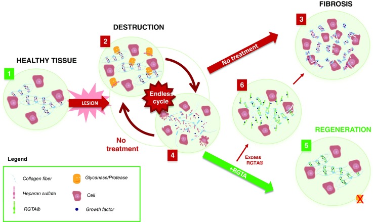
The importance of extracellular matrix (ECM) integrity in maintaining normal tissue function is highlighted by numerous pathologies and situations of acute and chronic injury associated with dysregulation or destruction of ECM components. Heparan sulfate (HS) is a key component of the ECM, where it fulfils important functions associated with tissue homeostasis. Its degradation following tissue injury disrupts this delicate equilibrium and may impair the wound healing process. ReGeneraTing Agents (RGTA®s) are polysaccharides specifically designed to replace degraded HS in injured tissues. The unique properties of RGTA® (resistance to degradation, binding and protection of ECM structural and signaling proteins, like HS) permit the reconstruction of the ECM, restoring both structural and biochemical functions to this essential substrate, and facilitating the processes of tissue repair and regeneration. Here, we review 25 years of research surrounding this HS mimic, supporting the mode of action, pre-clinical studies and therapeutic efficacy of RGTA® in the clinic, and discuss the potential of RGTA® in new branches of regenerative medicine.

Keywords: Extracellular scaffold; Heparan sulfate mimics; RGTA®; Regeneration.


Conflict of interest statement

Conflicts of interest

Denis Barritault is co-inventor and co-owner in the patents describing the RGTA® technology and president of the company OTR3, manufacturer of RGTA®-based products. None of the other authors have commercial interests.

Ethical approval

This review article does not contain novel studies with human participants or animals performed by any of the authors.

Figures

Fig. 1
Fig. 1
Mode of Action of ReGeneraTing Agent (RGTA®) Following Extensive Tissue Damage. 1. The Extracellular Matrix (ECM) organization in healthy tissue (left panel). Blue dots correspond to communication peptides, stored and protected by glycosaminoglycans. Heparan sulfate (HS) (light pink helices) links the matrix proteins (white fibers) to form a scaffold around cells. 2. Following extensive tissue damage, HS is degraded by glycanase (yellow figurines), the ECM scaffold structure is destroyed and proteases (yellow figurines) may enter, destroying matrix proteins and stored growth factors. 3. A rapid repair response is triggered resulting in scars or fibroses. 4. If the cause of the lesion is not resolved, wounds remain in an inflammatory phase, leading to a cycle of repair and destruction. 5. RGTA® (green helices) replaces degraded HS, allowing reconstruction of the ECM scaffold and restoring the cellular microenvironment. This facilitates tissue regeneration and restoration of tissue to a state similar to that of healthy tissue. 6. Excess RGTA® may hinder the healing process by drawing growth factors and communication peptides away from the wound site. In this situation, the bioavailability of growth factors required for efficient wound healing is reduced, emphasizing the need to optimize the RGTA® treatment protocol (dose and timing) in accordance with the tissue specific matrix turnover
Fig. 2
Fig. 2
Filling of craniotomy defects. A. Operative view of the defect. X-ray of defects treated with a collagen plaster soaked in 30 ul of B. saline, C. RGTA® solution (25 μg/ml) D. RGTA® solution (100 μg/ml) and E. RGTA® solution (400 ug/ml), after 4 weeks. F. Full macroscopic view of the skull defect with top arrows showing both sides of the defect and bottom arrows indicating new bone formation. Note the convex shape of the new bone structure, matching with expected original bone. v at the center indicates the saggital vein. G. Enlargement showing the lamellar organization of the flat bone as expected from a skull. H. alkaline phosphatase positive osteoblastic cells

References

    1. Sarrazin S, Lamanna WC, Esko JD. Heparan sulfate proteoglycans. Cold Spring Harb. Perspect. Biol. 2011;3(7):a004952. doi: 10.1101/cshperspect.a004952. - DOI - PMC - PubMed
    1. Hynes RO, Naba A. Overview of the matrisome--an inventory of extracellular matrix constituents and functions. Cold Spring Harb. Perspect. Biol. 2012;4(1):a004903. doi: 10.1101/cshperspect.a004903. - DOI - PMC - PubMed
    1. Bonnans C, Chou J, Werb Z. Remodelling the extracellular matrix in development and disease. Nat. Rev. Mol. Cell Biol. 2014;15(12):786–801. doi: 10.1038/nrm3904. - DOI - PMC - PubMed
    1. Cox TR, Erler JT. Remodeling and homeostasis of the extracellular matrix: implications for fibrotic diseases and cancer. Dis. Model. Mech. 2011;4(2):165–78. doi: 10.1242/dmm.004077. - DOI - PMC - PubMed
    1. Gallagher JT. Multiprotein signalling complexes: regional assembly on heparan sulphate. Biochem. Soc. Trans. 2006;34(Pt 3):438–41. doi: 10.1042/BST0340438. - DOI - PubMed

MeSH terms