. 2017 Nov;30(22):2734-2741.
doi: 10.1080/14767058.2016.1261400. Epub 2016 Dec 7.

Caffeine combined with sedative/anesthetic drugs triggers widespread neuroapoptosis in a mouse model of prematurity

Affiliations

Caffeine combined with sedative/anesthetic drugs triggers widespread neuroapoptosis in a mouse model of prematurity

Omar Hoseá Cabrera et al. J Matern Fetal Neonatal Med. 2017 Nov.

Abstract

Objectives: Caffeine (CAF) and sedative/anesthetic drugs (SADs) are often coadministered to premature infants in the neonatal intensive care unit (NICU). While SAD neurotoxicity in the developing brain is well established, it is not fully clear whether CAF interacts with SADs and whether this interaction is detrimental. Using a mouse model of prematurity, we hypothesized that CAF would increase apoptotic neurotoxicity when coadministered with SADs.

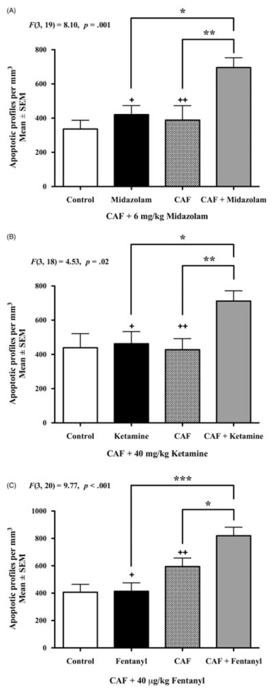
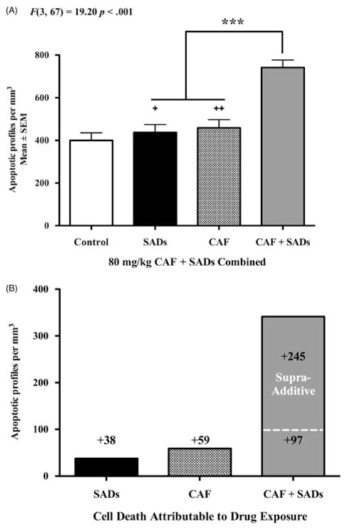
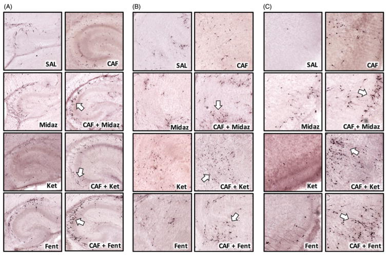
Methods: Postnatal day 3 mice were treated with vehicle or 80 mg/kg CAF prior to challenge with 6 mg/kg midazolam, 40 mg/kg ketamine, or 40 μg/kg fentanyl. Six hours later, pups were sacrificed for activated caspase 3 (AC3) immunohistochemistry, and number of AC3 positive cells per mm3 throughout neocortex, hippocampus, caudate, thalamus, and colliculi was analyzed.

Results: CAF caused a statistically significant increase in AC3 positive cells when coadministered with midazolam (p = 0.002), ketamine (p = 0.014), or fentanyl (p < 0.001). Our composite dataset suggests that the addition of CAF to these SADs has a supra-additive effect, causing more neurotoxicity than expected.

Conclusions: CAF may augment the neurotoxic action of SADs indicated for neonatal sedation/anesthesia in the NICU by triggering widespread apoptosis in the developing brains of premature infants.

Keywords: Caffeine; apoptosis; fentanyl; ketamine; midazolam; premature infant.


Conflict of interest statement

Disclosure statement

The authors report no conflicts of interest. The authors alone are responsible for the content and writing of this article.

Figures

Figure 1
Figure 1. Apoptogenic action of CAF with NICU SADs
Six hours after drug administration, PND3 brains were assessed for apoptotic neurotoxicity via AC3 immunolabeling (n ≥5 per group). Cell counts for the control groups represent physiologic apoptosis, that is, the background rate of natural cell death in the PND3 brain. Panel A displays the mean density counts of apoptotic profiles after challenge with CAF + 6 mg/kg midazolam. A single exposure to midazolam or CAF caused no increase in apoptosis compared to controls. However, when CAF was combined with midazolam there was a statistically significant increase in apoptosis compared to either alone. Panel B demonstrates the neurotoxic reaction of CAF +40 mg/kg ketamine. The CAF + ketamine cocktail caused a significant increase in apoptotic profiles per mm3 compared to CAF alone or ketamine alone. The apoptotic neurotoxicity of CAF + 40 μg/kg fentanyl is illustrated in Panel C. CAF + fentanyl coexposure led to a statistically significant increase in mean number of apoptotic cells per mm3 versus the CAF only and fentanyl only groups. Panel A: +Cohen’s d = 2.56, ++Cohen’s d = 2.72 compared to CAF + midazolam. Panel B: +Cohen’s d = 1.45, ++Cohen’s d = 1.75 compared to CAF + ketamine. Panel C: +Cohen’s d = 2.47, ++Cohen’s d = 1.23 compared to CAF +Fentanyl *p <0.05, **p <0.01, ***p <0.001.
Figure 2
Figure 2
Overview of the apoptogenic action of CAF. To provide a general overview of the pro-apoptotic action CAF when combined with SADs, we generated a composite dataset (Panel A) of the density counts for all animals exposed to SADs (midazolam, ketamine, or fentanyl), CAF, or CAF + SADs at PND3. In this composite dataset, the addition of CAF to the SADs tested caused significantly more apoptotic neurotoxicity than either CAF or SAD alone. Panel B displays the composite dataset from Panel A adjusted to reflect cell death attributable to drug exposure. By subtracting out the control value representing the natural background rate of physiologic apoptosis, the remaining values reflect the amount of apoptotic cell death attributable to drug exposure. Exposure to SADs added 38 AC3-positive profiles per mm3, while exposure to CAF added 59 AC3-positive profiles per mm3. The dashed line is the number of apoptotic cells that would be expected by simply adding together SAD apoptosis and CAF apoptosis (38 + 59 = 97). However, the mean number of AC3-positive profiles in the CAF + SAD group was much greater than predicted, resulting in an additional 245 apoptotic cells per mm3. Our data suggest that CAF may have supra-additive properties when combined with SADs. Panel A: +Cohen’s d= 2.10, ++Cohen’s d = 1.66 compared to CAF + SADs ***p <0.001.
Figure 3
Figure 3
Representative images of CAF + SAD neurotoxicity in the PND3 mouse brain. The addition of CAF to SADs caused apoptotic neurotoxicity in every region examined. The above panels are representative images of select neural substrates key to learn and memory and information processing: hippocampus, thalamus, and retrosplenial cortex. Each CAF + SAD pairing produced a unique pattern of cell death in those regions, and generally, many cells were in an advanced state of degeneration (white arrows) characteristic of late-stage apoptosis. Note the diverse morphology of AC3-positive cells, suggesting that a specific cell type is not preferentially vulnerable to CAF + SAD neurotoxicity. Panel A illustrates apoptotic neurotoxicity in the hippocampus after CAF + SAD challenge. Numerous apoptotic profiles are visible and scattered throughout the CA2 and CA3 hippocampal fields. Panel B focuses on anterior thalamus and neighboring dorsal and ventral thalamic nuclei. In control animals, few apoptotic cells were observed in these regions. The CAF + SAD cocktail substantially increased the density of AC3-positive profiles, many of which possess a medium-sized stellate morphology. Panel C represents detailed views of neuroapoptosis in retrosplenial cortex. These images reveal that CAF combined with midazolam, ketamine, or fentanyl caused a robust apoptotic reaction that involved small pyramidal neurons in superficial cortical layers and larger projection neurons in deeper cortical layers.

References

    1. Jevtovic-Todorovic V, Absalom AR, Blomgren K, et al. Anaesthetic neurotoxicity and neuroplasticity: an expert group report and statement based on the BJA Salzburg Seminar. Br J Anaesth. 2013;111:143–51. - PMC - PubMed
    1. Filan PM, Hunt RW, Anderson PJ, et al. Neurologic outcomes in very preterm infants undergoing surgery. J Pediatr. 2012;160:409–14. - PubMed
    1. Morriss FH, Jr, Saha S, Bell EF, et al. Eunice Kennedy Shriver National Institute of Child H, Human Development Neonatal Research N. Surgery and neurodevelopmental outcome of very low-birth-weight infants. JAMA Pediatr. 2014;168:746–54. - PMC - PubMed
    1. Henderson-Smart DJ, Steer P. Methylxanthine treatment for apnea in preterm infants. Cochrane Database Syst Rev. 2001:CD000140. - PubMed
    1. Schmidt B, Roberts RS, Davis P, et al. Caffeine for Apnea of Prematurity Trial G. Caffeine therapy for apnea of prematurity. N Engl J Med. 2006;354:2112–21. - PubMed

MeSH terms