

P-wave dispersion: an update
- PMID: 27924760
- PMCID: PMC5197451
- DOI: 10.1016/j.ipej.2016.10.002
P-wave dispersion: an update
Abstract
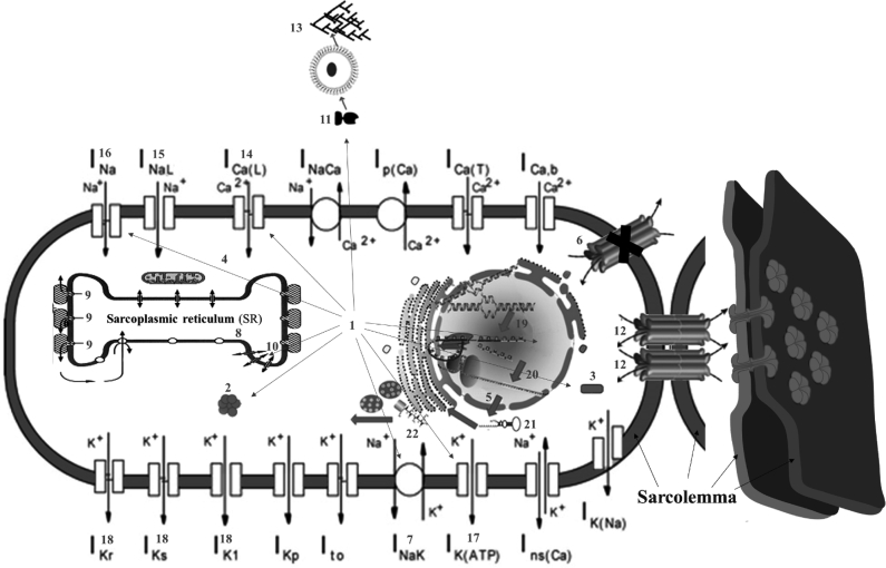
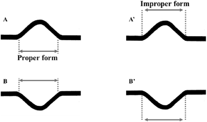
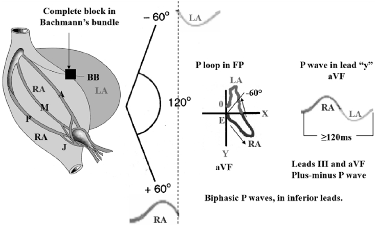
P-wave dispersion (PWD, Pd or Pdis) is a noninvasive electrocardiographic (ECG) marker for atrial remodeling and predictor for atrial fibrillation (AF). PWD is defined as the difference between the widest and the narrowest P-wave duration recorded from the 12 ECG leads. Increased P-wave duration and PWD reflect prolongation of intraatrial and interatrial conduction time with lack of a well-coordinated conduction system within the atrial muscles, with inhomogeneous, asynchronic, pro-inflammatory and anti-inflammatory effect mediated by interleukin-6 (IL-6) in patients with the CG + GG genotype IL-6 -634C/G polymorphism [1] and discontinuous propagation of sinus impulses mainly between the left and right atria, interstitial/extracellular fibroblast activation and collagen deposition with fibrosis (via TGF-β) in atrial tissue, insufficient blood supply, significant not isotropic myoelectric activity, and thin wall thickness and consequent expansion tendency all well-known electrophysiological characteristics in patients with atrial arrhythmias and especially paroxysmal atrial fibrillation (PAF) [2].
Keywords: Interatrial block; Intraatrial block; P-wave dispersion; P-wave duration; Paroxysmal atrial fibrillation.
Copyright © 2016 Indian Heart Rhythm Society. Production and hosting by Elsevier B.V. All rights reserved.
Figures
References
-
- Justo F., Fuller H., Nearing B.D., Rajamani S., Belardinelli L., Verrier R.L. Inhibition of the cardiac late Na+ current with eleclazine protects against ischemia-induced vulnerability to atrial fibrillation and reduces atrial and ventricular repolarization abnormalities in the absence and presence of concurrent adrenergic stimulation. Heart Rhythm. 2016 S1547–5271(16)30438-6. (in press) - PubMed
-
- Aytemir K., Ozer N., Atalar E. P wave dispersion on 12-lead electrocardiography in patients with paroxysmal atrial fibrillation. Pacing Clin Electrophysiol. 2000;23(7):1109–1112. - PubMed
Publication types
LinkOut - more resources
Full Text Sources
Other Literature Sources
