. 2016 Dec 7;8(4):251-264.
doi: 10.1002/cpch.14.

Cyclic Immunofluorescence (CycIF), A Highly Multiplexed Method for Single-cell Imaging

Affiliations

Cyclic Immunofluorescence (CycIF), A Highly Multiplexed Method for Single-cell Imaging

Jia-Ren Lin et al. Curr Protoc Chem Biol. .

Abstract

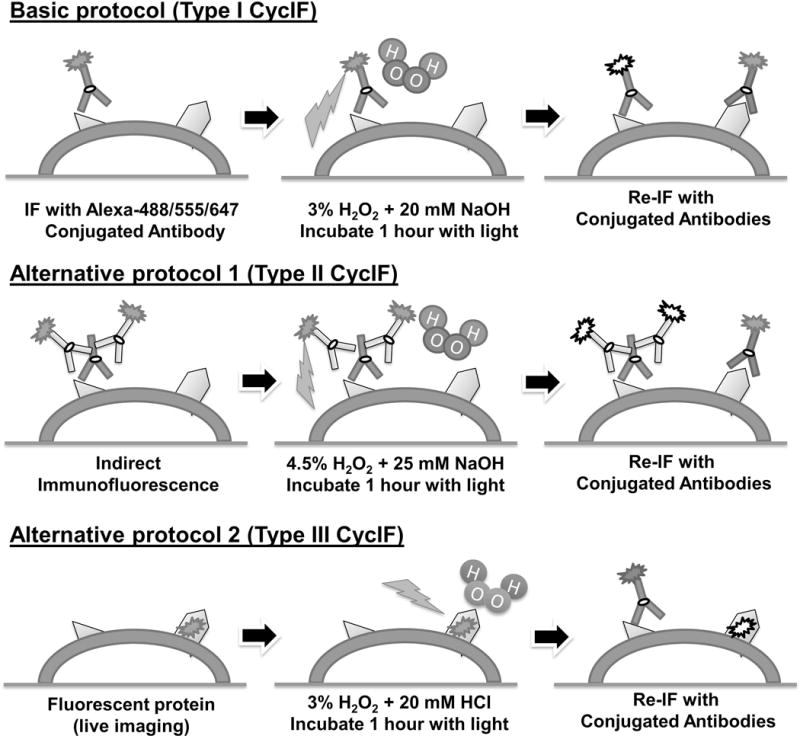
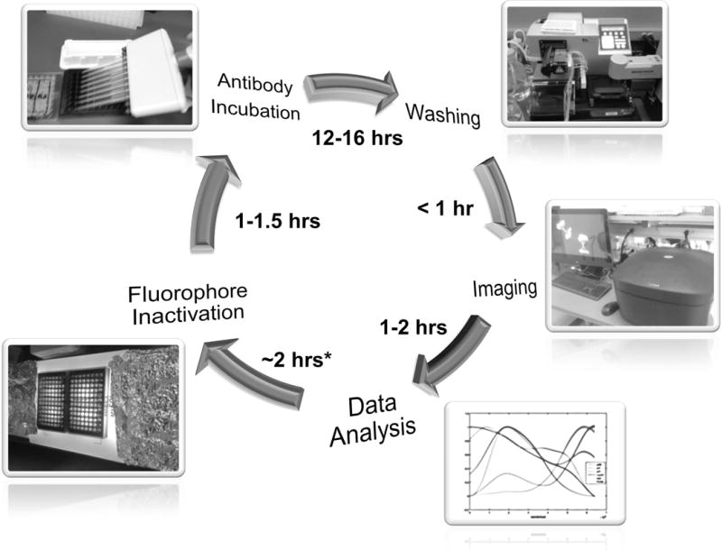
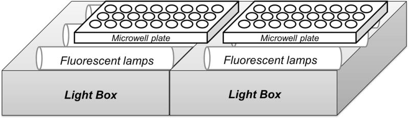
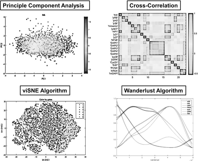
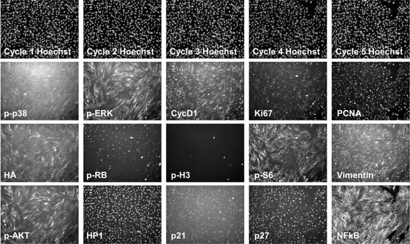
Cyclic Immunofluorescence (CycIF) is a public-domain method for performing highly multiplexed immunofluorescence imaging using a conventional epifluorescence microscope. It uses simple reagents and existing antibodies to construct images with up to 30 channels by sequential 4- to 6-channel imaging followed by fluorophore inactivation. Three variant methods are described, the most generally useful of which involves staining fixed cells with antibodies directly conjugated to Alexa Fluor dyes and imaging in four colors, inactivating fluorophores using a mild base in the presence of hydrogen peroxide and light, and then performing another round of staining and imaging. Cell morphology is preserved through multiple rounds of CycIF, and signal-to-noise ratios appear to increase. Unlike antibody-stripping methods, CycIF is gentle and optimized for monolayers of cultured cells. A second protocol involves indirect immunofluorescence and a third enables chemical inactivation of genetically encoded fluorescent proteins, allowing multiplex immunofluorescence to be combined with live-cell analysis of cells expressing fluorescent reporter proteins. © 2016 by John Wiley & Sons, Inc.

Keywords: CycIF; high-content imaging; immunofluorescence; multiplexing; systems biology.


Figures

Figure 1
Figure 1. Three ways to achieve highly multiplexed read-outs in single cells
See text for details.
Figure 2
Figure 2. Experimental workflow with time estimations
Illustrated here is a typical CycIF cycle. Start from the antibody incubation step and follow by washing, imaging and preliminary data analysis. Fluorophore inactivation is performed after the confirmation of image/data quality. One full cycle can be completed in 24 hours.
Figure 3
Figure 3
The light device for fluorophore inactivation.
Figure 4
Figure 4. Analysis of high-dimensional CycIF data
Shown here are four methods for high-dimensional data analysis of CycIF data. Principal Component Analysis (PCA) and viSNE algorithms are generally used for dimensionality reduction and visualization of CycIF data (Amir et al., 2013; Lin et al., 2015). The cross-correlation method is used for probing the relationships between different signals measured by CycIF. The Wanderlust algorithm is a graph-based trajectory detection method, mapping the high-dimensional single-cell CycIF data to a one-dimensional path based on most likely transitional cell states (Bendall et al., 2014; Lin et al., 2015).
Figure 5
Figure 5. Example images for a five-round CycIF experiment
Retinal pigment epithelium (RPE) cells were cultured and fixed in 96-well plates. Five rounds of CycIF staining were performed and representative images are shown with indicated antibodies.

References

    1. Amir ED, Davis KL, Tadmor MD, Simonds EF, Levine JH, Bendall SC, Pe’er D. viSNE enables visualization of high dimensional single-cell data and reveals phenotypic heterogeneity of leukemia. Nature Biotechnology. 2013;31(6):545–52. doi: 10.1038/nbt.2594. - DOI - PMC - PubMed
    1. Angelo M, Bendall SC, Finck R, Hale MB, Hitzman C, Borowsky AD, Nolan GP. Multiplexed ion beam imaging of human breast tumors. Nature Medicine. 2014;20(4):436–442. doi: 10.1038/nm.3488. - DOI - PMC - PubMed
    1. Bendall SC, Davis KL, Amir EAD, Tadmor MD, Simonds EF, Chen TJ, Pe’er D. Single-cell trajectory detection uncovers progression and regulatory coordination in human B cell development. Cell. 2014;157(3):714–25. doi: 10.1016/j.cell.2014.04.005. - DOI - PMC - PubMed
    1. Donaldson JG. Immunofluorescence staining. Current Protocols in Immunology/Edited by John E Coligan … [et Al], Chapter 21. 2002 doi: 10.1002/0471142735.im2103s48. Unit 21.3. - DOI - PubMed
    1. Frei AP, Bava FA, Zunder ER, Hsieh EWY, Chen SY, Nolan GP, Gherardini PF. Highly multiplexed simultaneous detection of RNAs and proteins in single cells. Nature Methods. 2016;13(3):269–275. doi: 10.1038/nmeth.3742. - DOI - PMC - PubMed