. 2016 Dec;186(6):624-637.
doi: 10.1667/RR14569.1. Epub 2016 Dec 7.

Modeling Heavy-Ion Impairment of Hippocampal Neurogenesis after Acute and Fractionated Irradiation

Affiliations

Modeling Heavy-Ion Impairment of Hippocampal Neurogenesis after Acute and Fractionated Irradiation

Eliedonna Cacao et al. Radiat Res. 2016 Dec.

Abstract

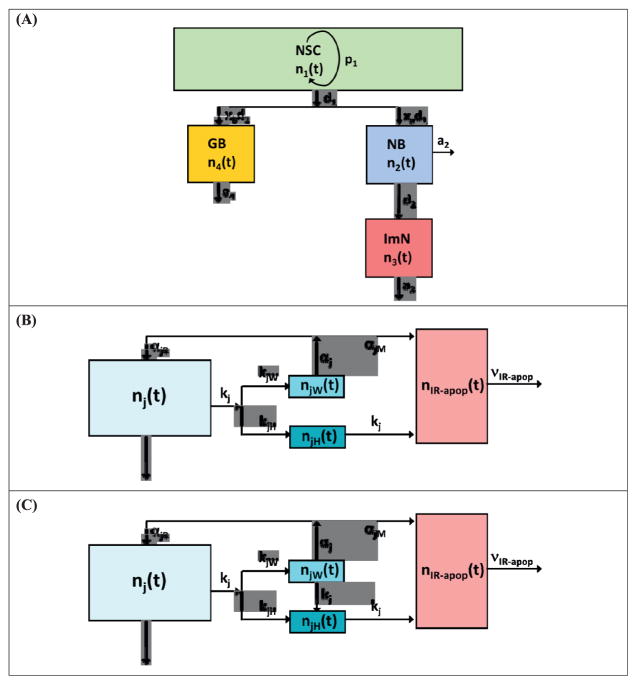
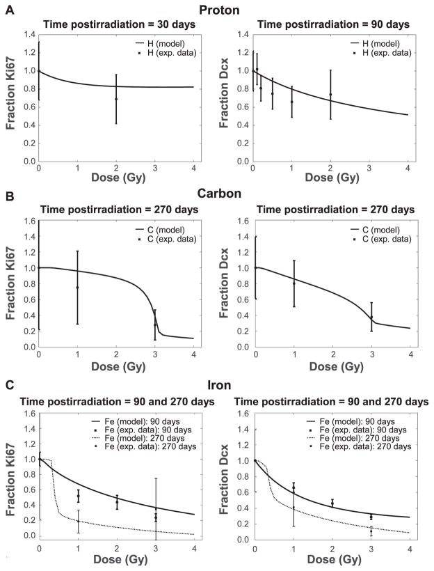
Radiation-induced impairment of neurogenesis in the hippocampal dentate gyrus is a concern due to its reported association with cognitive detriments after radiotherapy for brain cancers and the possible risks to astronauts chronically exposed to space radiation. Here, we have extended our recent work in a mouse model of impaired neurogenesis after exposure to low-linear energy transfer (LET) radiation to heavy ion irradiation. To our knowledge, this is the first report of a predictive mathematical model of radiation-induced changes to neurogenesis for a variety of radiation types after acute or fractionated irradiation. We used a system of nonlinear ordinary differential equations (ODEs) to represent age, time after exposure and dose-dependent changes to several cell populations participating in neurogenesis, as reported in mouse experiments. We considered four compartments to model hippocampal neurogenesis and, consequently, the effects of radiation in altering neurogenesis: 1. neural stem cells (NSCs); 2. neuronal progenitor cells or neuroblasts (NB); 3. immature neurons (ImN); and 4. glioblasts (GB), with additional consideration of microglial activation. The model describes the negative feedback regulation on early and late neuronal proliferation after irradiation, and the dynamics of the age dependence of neurogenesis. We compared our model to experimental data for X rays, and protons, carbon and iron particles, including data for fractionated iron-particle irradiation. Heavy-ion irradiation is predicted to lead to poor recovery or no recovery from impaired neurogenesis at doses as low as 0.5 Gy in mice. This is only partially ameliorated by dose fractionation, which suggests important implications for Hardon therapy near the Bragg peak, and possibly for space radiation exposures as well. Predictions of the threshold doses where neurogenesis recovery fails for given radiation types are described, and the role of subthreshold transient impairments are briefly discussed.


Figures

FIG. 1
FIG. 1
Hippocampal neurogenesis model. Panel A: Schematic diagram of neurogenesis model showing cell population, nj, where j = 1–4, represents neural stem cells [NSCs (n1)], neuroblasts [NBs (n2)], immature neurons [ImNs (n3)] and glioblasts [GBs (n4)]. Panels B and C: Neurogenesis model after irradiation. Apoptotic cell population due to exposure is indicated by nIRapop. Radiation damages are described by rate constants kj, where kj = kjW + kjH. Damage repair and misrepair rates are represented by αjR and αjM, respectively, where αj = αjR + αjM. Panel B: The fate of weakly damaged cells (njW) is to undergo either repair or apoptosis (misrepaired). Panel C: A fraction of weakly damaged cells is converted to heavily damaged cells (njH).
FIG. 2
FIG. 2
Age-dependent neuronal cell population of mouse hippocampal neurogenesis. Modeling dynamics of neuronal cell population includes NSCs, NBs and ImNs, described as surviving fraction with corresponding experimental (exp.) data (panel A) (21, 37) or cell population with comparison of mouse and human life stages (panel B) (38).
FIG. 3
FIG. 3
Panels A–C: Dose-dependent response of hippocampal neurogenesis to acute exposure of proton, carbon or iron radiation, respectively. Neurogenesis is evaluated using proliferation marker Ki67 (left side) and ImN marker Dcx (right side) at a specified postirradiation time, in comparison to experimental data [protons (H) (16), carbon (C) (25) and iron (Fe) (24, 25)].
FIG. 4
FIG. 4
Newly born activated microglia and change in neurogenic fate at 60 days postirradiation. Panel A: Fractional increase in newly born activated microglia (left side) and fold decrease in neurogenic fate of newly born cells (right side) after acute exposure of iron radiation [model vs. experimental results (27)]. Panel B: Different particle radiation (X ray/proton vs. carbon vs. iron) effects on fractional increase in newly born activated microglia (left side) and fold decrease in neurogenic fate of newly born cells (right side).
FIG. 5
FIG. 5
Panels A–D: Modeling dynamics of hippocampal neurogenesis, represented by fraction of Ki67 (left side) and Dcx (right side), after acute exposure to different doses of X-ray, proton, carbon and iron radiation, respectively.
FIG. 6
FIG. 6
Threshold doses for immature neurons exposed to different radiation types. Threshold dose, defined as the minimum dose of radiation where recovery does not occur by 270 days postirradiation, is plotted against the characteristic dose for damage of immature neurons (D03).
FIG. 7
FIG. 7
Acute and fractionated exposure to different radiation types. Panel A: Neurogenesis is evaluated using immature neuron marker Dcx after acute (1 Gy) or fractionated (5 × 0.20 Gy/day) exposure to proton, carbon or iron radiation at 1 (left) and 90 days postirradiation. [experimental results for iron irradiation are from ref. (29)]. Panels B–D: Effects of higher dose of acute (5 Gy) or fractionated (5 × 1 Gy/day) radiation exposure on proliferation marker Ki67 (left side) and Dcx (right side) at 60, 90 and 270 days postirradiation, respectively.

References

    1. Wefel JS, Vardy J, Ahles T, Schagen SB. International Cognition and Cancer Task Force recommendations to harmonise studies of cognitive function in patients with cancer. Lancet Onc. 2011;12:703–8. - PubMed
    1. Dias GP, Hollywood R, Bevilaqua MCN, da Luz ACD, Hindges R, Nardi AE, et al. Consequences of cancer treatments on adult hippocampal neurogenesis: implications for cognitive function and depression symptoms. Neuro-Onc. 2014;0:1–17. - PMC - PubMed
    1. Greene-Schloesser D, Robbins ME, Peiffer AM, Shaw EG, Wheeler KT, Chan MD. Radiation-induced brain injury: A review. Front Oncol. 2012;2:73. - PMC - PubMed
    1. Monje ML, Palmer T. Radiation injury and neurogenesis. Curr Opin Neurol. 2003;16:129–34. - PubMed
    1. Monje ML, Vogel H, Masek M, Ligon KL, Fischer PG, Palmer TD. Impaired human hippocampal neurogenesis after treatment for central nervous system malignancies. Annals Neurology. 2007;62:515–20. - PubMed