. 2017 Jan 10;8(2):2719-2730.
doi: 10.18632/oncotarget.13739.

Macrophage migration inhibitory factor activates inflammatory responses of astrocytes through interaction with CD74 receptor

Affiliations

Macrophage migration inhibitory factor activates inflammatory responses of astrocytes through interaction with CD74 receptor

Yu Su et al. Oncotarget. .

Abstract

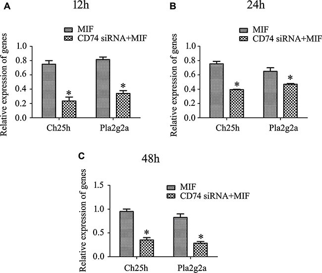
Astrocytes, the major glial cell population of the central nervous system (CNS), play important physiological roles related to CNS homeostasis. Growing evidence demonstrates that astrocytes trigger innate immune responses under challenge of a variety of proinflammatory cytokines. Macrophage migration inhibitory factor (MIF), a proinflammatory cytokine mainly secreted from monocytes/macrophages, is involved in inflammation-associated pathophysiology. Here, we displayed that expression of MIF significantly increased following spinal cord injury, in colocalization with microglia and astrocytes. MIF elicited inflammatory responses of astrocytes via activation of CD74 receptor and extracellular signal-related kinase (ERK) pathway. Transcriptome analysis revealed that inflammation-related factors cholesterol 25-hydroxylase (Ch25h) and phospholipase A2-IIA (Pla2g2a), downstream of MIF/CD74 axis, were potentially implicated in the mediating inflammatory response of astrocytes. Our results provided a new target for interference of CNS inflammation after insults.

Keywords: CD74; MIF; astrocyte; inflammation; spinal cord.


Conflict of interest statement

CONFLICTS OF INTEREST

The authors have declared that no competing interests exist.

Figures

Figure 1
Figure 1. Expression of MIF increased following spinal cord injury
(A) Western blot analysis of MIF expression following spinal cord contusion at 0d, 1d, 4d and 7d, respectively. (B, C) Immunostaining showed colocalization of MIF with GFAP- and Iba-1- positive cells. Rectangle indicates region magnified below. Quantities were normalized to endogenous β-actin. Error bars represent the standard deviation (P < 0.05). Scale bars, 100 μm.
Figure 2
Figure 2. Examination of inflammatory activation in astrocytes following stimuli with gradient recombinant MIF
(A) Showing purified primary astrocytes stained with GFAP and Hoechst 33342. (B) Expression analysis of TNFα and IL-1β by RT-PCR, following astrocytes treatment with 0–2.5 μg/ml recombinant MIF for 24 h. (C) Cell supernatants and lysates were tested by ELISA for the cytokines TNFα and IL-1β. (D) Western blot analysis of p65NFκB activation in astrocytes. Quantities were normalized to endogenous β-actin. Error bars represent the standard deviation (P < 0.05). Scale bars, 50 μm.
Figure 3
Figure 3. Binding assay of MIF with CD74 receptor in the primary astrocytes
Immunoprecipitation using anti-MIF or -CD74 antibody and detection of the components of the MIF- or CD74-associated complexes with anti-CD74 or -MIF antibody.
Figure 4
Figure 4. Knockdown of C74 receptor affected MIF-induced inflammatory responses in astrocytes
(A) Determination of siRNA transfection efficiency by Cy3 control. (B) Interference efficiency of three siRNA oligonucleotides for CD74 was measured by RT-PCR, and siRNA2 was used for the knockdown experiments. (C, D) Expression analysis of TNFα and IL-1β by RT-PCR, following astrocytes treated with siRNA2 oligonucleotides or scramble for 48 h, and then with 2 μg/ml recombinant MIF for 24 h. (E, F) Western blot analysis of pERK and p65NFκB following astrocytes treated with siRNA2 oligonucleotides or scramble for 48 h, and then with 2 μg/ml recombinant MIF for 15 min, 30 min and 60 min, respectively. Quantities were normalized to endogenous β-actin. Error bars represent the standard deviation (P < 0.05). Scale bars, 10 μm.
Figure 5
Figure 5. MIF stimulates astrocytes proliferation in vitro
(A) Effects of MIF on proliferation of primary cultured astrocytes. (B) Quantification data as shown in (A). Error bars represent the standard deviation. *P < 0.05. Scale bars, 100 μm.
Figure 6
Figure 6. Functional annotations of DEGs in the astrocytes following knockdown of CD74
(A), (C) and (E) Bar graphs of DEGs following knockdown of CD74 for 48 h, and then treated with 2 μg/ml recombinant MIF at 12 h, 24 h and 48 h, respectively. (B), (D) and (F) Most significantly enriched groups for the DEGs relating to pathways.
Figure 7
Figure 7. Expression profiling of integrated DEGs in the astrocytes following knockdown of CD74
(A) Integration of DEGs at 12 h, 24 h and 48 h. (B) Most significantly enriched groups for the integrated DEGs relating to pathways. (C) Heatmap and cluster dendrogram of integrated DEGs.
Figure 8
Figure 8. A reconstructed gene network was created using the Ingenuity Pathway Analysis Software (IPA) on the basis of integrated DEGs
Figure 9
Figure 9
RT-PCR analysis of Ch25h and Pla2g2a expression in primary astrocytes following knockdown of CD74 for 48 h, and then treated with 2 μg/ml recombinant MIF at 12 h (A), 24 h (B) and 48 h (C), respectively.

References

    1. Ransohoff RM, Brown MA. Innate immunity in the central nervous system. J Clin Invest. 2012;122:1164–1171. - PMC - PubMed
    1. McKimmie CS, Graham GJ. Astrocytes modulate the chemokine network in a pathogen-specific manner. Biochem Biophys Res Commun. 2010;394:1006–1011. - PubMed
    1. Panenka W, Jijon H, Herx LM, Armstrong JN, Feighan D, Wei T, Yong VW, Ransohoff RM, MacVicar BA. P2X7-like receptor activation in astrocytes increases chemokine monocyte chemoattractant protein-1 expression via mitogen-activated protein kinase. J Neurosci. 2001;21:7135–7142. - PMC - PubMed
    1. Stoll G, Jander S, Schroeter M. Inflammation and glial responses in ischemic brain lesions. Prog Neurobiol. 1998;56:149–171. - PubMed
    1. Glabinski AR, Balasingam V, Tani M, Kunkel SL, Strieter RM, Yong VW, Ransohoff RM. Chemokine monocyte chemoattractant protein-1 is expressed by astrocytes after mechanical injury to the brain. J Immunol. 1996;156:4363–4368. - PubMed

MeSH terms

Substances

LinkOut - more resources