. 2017 Mar 1;312(3):C233-C243.
doi: 10.1152/ajpcell.00151.2016. Epub 2016 Dec 7.

Prenatal myonuclei play a crucial role in skeletal muscle hypertrophy in rodents

Affiliations

Prenatal myonuclei play a crucial role in skeletal muscle hypertrophy in rodents

Fuminori Kawano et al. Am J Physiol Cell Physiol. .

Abstract

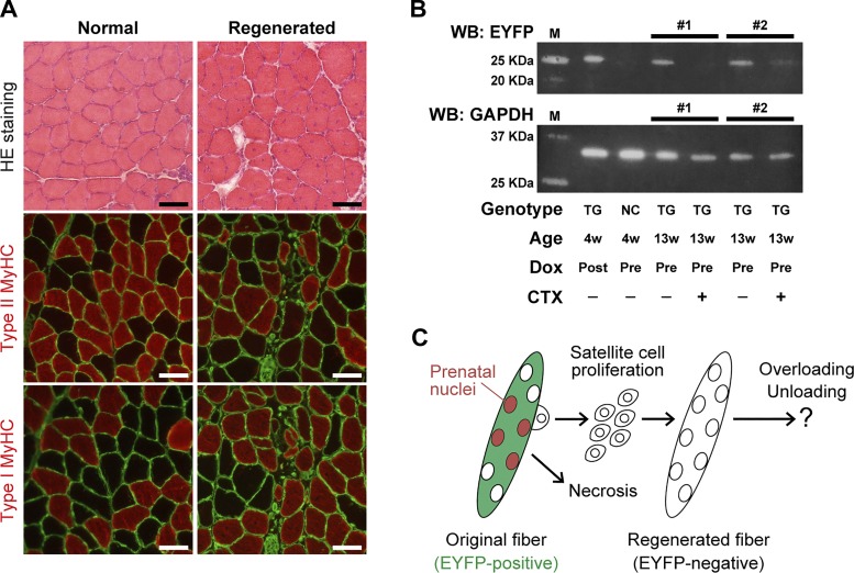
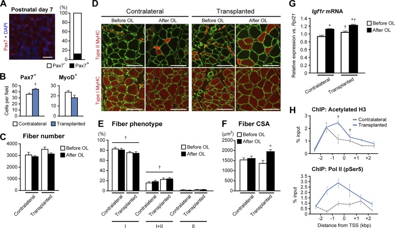
Multinucleated muscle fibers are formed by the fusion of myogenic progenitor cells during embryonic and fetal myogenesis. However, the role of prenatally incorporated myonuclei in the skeletal muscle fibers of adult animals is poorly understood. We demonstrated, using muscle-specific reporter mice, that the prenatal myonuclei remained in the adult soleus muscle, although cardiotoxin injection caused the loss of prenatal myonuclei. Overloading by the tendon transection of synergists failed to induce compensatory hypertrophy in regenerated soleus muscle fibers of adult rats, whereas unloading by tail suspension normally induced the fiber atrophy. Loss of hypertrophying function correlated with the lowered histone acetylation at the transcription start site of Igf1r gene, which was one of the genes that did not respond to the overloading. These parameters were improved by the transplantation of cells harvested from the juvenile soleus muscles of neonatal rats in association with enhanced histone acetylation of Igf1r gene. These results indicated that the presence of prenatal myonuclei was closely related to the status of histone acetylation, which could regulate the responsiveness of muscle fibers to physiological stimuli.

Keywords: histone modification; injury; myonucleus; regeneration; skeletal muscle fiber.


Figures

Fig. 1.
Fig. 1.
Conditional expression of EYFP in prenatal myonuclei of transgenic mice. A: H&E staining (top row) and immunohistochemistry for type II (red in middle row) or type I (red in bottom row) MyHC and laminin (green) in normal (right leg) and regenerated (left leg) soleus muscles. Scale bar, 50 µm. B: EYFP and GAPDH expressions were detected by Western blotting in soleus muscles of noncarrier (NC, HSA-rtTA/TRE-Cre−/−; R26R-YFP+/+) and transgenic (TG, HSA-rtTA/TRE-Cre+/−; R26R-YFP+/+) mice at the age of 4 wk. Doxycycline (Dox) was given in the drinking water during the prenatal period (Pre) via the pregnant mice or constantly after birth (Post). M indicates the molecular weight marker, and #1 or #2 indicates the right (CTX–) and left (CTX+) muscles sampled from the same mouse. C: model of the remaining and loss of the prenatal nuclei in the muscle fiber. Note that prenatally incorporated nuclei (indicated in red) remain in the adult muscle fiber (EYFP-positive). These nuclei are lost after the muscle injury, followed by the subsequent formation of new fiber (EYFP-negative) by the proliferation of satellite cells.
Fig. 2.
Fig. 2.
Lack of hypertrophying function in regenerated fibers. A: total fiber numbers in whole cross section of normal and regenerated soleus muscles before (open bars) and after (closed bars) overloading (OL). B: immunohistochemically stained sections labeled by type II MyHC (red in top row) or type I MyHC (red in bottom row) and laminin (green) in normal and regenerated soleus muscles of rats before and after OL. Scale bar, 100 µm. C: distribution of fibers expressing type I, I+II, and II MyHC in normal and regenerated muscles before and after OL. D: responses of fiber CSA to OL. E: distribution of fiber CSA before (blue and red lines) vs. after (green and purple lines) OL in normal (panel at left) and regenerated (panel at right) muscles. F: changes in the expression of genes upregulated by OL in normal muscle determined using microarray. Numbers in parentheses indicate the number of probes in microarray. G: levels of Igf1r mRNA expression in normal and regenerated muscles before and after OL. H: distribution of acetylated H3 (panel at top) and Pol II (pSer5) (panel at bottom) near TSS, exon 2, and TTS of Igf1r gene determined by ChIP-qPCR. Experimental groups are indicated by the same colors as shown in E. *P < 0.05, before vs. after OL; †P < 0.05, normal vs. regenerated muscles.
Fig. 3.
Fig. 3.
Transplantation of developmental cells improved the responses of regenerated fibers to overloading. A: immunohistochemically stained sections labeled by Pax7 in the soleus muscle of neonatal rat at postnatal day 7 (panel at left). Percent distribution of Pax7-positive nuclei per total nuclei labeled by DAPI is shown (panel at right). B: numbers of Pax7- or MyoD-positive nuclei per field were counted in normal (white bars) and regenerated (blue bars) muscle after 8 wk of regeneration in adult rats. C: total fiber numbers in whole cross section of normal and regenerated soleus muscles before (open bars) and after (closed bars) overloading (OL). D: immunohistochemically stained sections labeled by type II MyHC (red in top row) or type I MyHC (red in bottom row) and laminin (green) in contralateral and transplanted soleus muscles of rats before and after OL. Scale bar, 100 µm. E: distribution of fibers expressing type I, I+II, and II MyHC in contralateral and transplanted muscles before and after OL. F: responses of fiber CSA to OL. G: levels of Igf1r mRNA expression in contralateral and transplanted muscles before and after OL. H: distribution of acetylated H3 (panel at top) and Pol II (pSer5) (panel at bottom) near TSS of Igf1r gene determined by ChIP-qPCR in contralateral (dashed lines) and transplanted (solid blue lines) muscles. *P < 0.05, before vs. after OL; †P < 0.05, contralateral vs. transplanted muscles. In E, †significant differences in contralateral and transplanted muscles identified by ANOVA.
Fig. 4.
Fig. 4.
Histone acetylation in quiescent satellite cells lying in adult soleus muscle. A: plots of sorted cells harvested from soleus muscles of adult rats. Panel at left shows the primary sorted cells (area enclosed by red line) negatively gated by CD31 and CD45 expression. Panel at right shows the result of the secondary sorting. Cells expressing both VCAM and CD29 (area enclosed by green line) were collected as quiescent satellite cells. Numbers shown in the plot areas indicate the percentage of cells distributed in each area. APC, allophycocyanin. B: distribution of acetylated H3 near TSS of Igf1r gene determined by ChIP-qPCR in donor soleus muscle (red) and satellite cells (black). See Fig. 2 for abbreviations.
Fig. 5.
Fig. 5.
Effects of hindlimb unloading in regenerated muscle fibers of adult rat soleus. A: immunohistochemically stained sections labeled by type II MyHC (red in top row) or type I MyHC (red in bottom row) and laminin (green) in normal and regenerated soleus muscles of rats before and after hindlimb unloading (HU). Scale bar, 100 µm. B: distribution of fibers expressing type I, I+II, and II MyHC in normal and regenerated muscles before (open bars) and after (closed bars) HU. C: responses of fiber CSA to HU. *P < 0.05, before vs. after HU; †P < 0.05, normal vs. regenerated muscles.

Similar articles

Cited by

References

    1. Allen DL, Linderman JK, Roy RR, Bigbee AJ, Grindeland RE, Mukku V, Edgerton VR. Apoptosis: a mechanism contributing to remodeling of skeletal muscle in response to hindlimb unweighting. Am J Physiol Cell Physiol 273: C579–C587, 1997. - PubMed
    1. Bajard L, Relaix F, Lagha M, Rocancourt D, Daubas P, Buckingham ME. A novel genetic hierarchy functions during hypaxial myogenesis: Pax3 directly activates Myf5 in muscle progenitor cells in the limb. Genes Dev 20: 2450–2464, 2006. doi:10.1101/gad.382806. - DOI - PMC - PubMed
    1. Beauchamp JR, Heslop L, Yu DS, Tajbakhsh S, Kelly RG, Wernig A, Buckingham ME, Partridge TA, Zammit PS. Expression of CD34 and Myf5 defines the majority of quiescent adult skeletal muscle satellite cells. J Cell Biol 151: 1221–1234, 2000. doi:10.1083/jcb.151.6.1221. - DOI - PMC - PubMed
    1. Biressi S, Bjornson CR, Carlig PM, Nishijo K, Keller C, Rando TA. Myf5 expression during fetal myogenesis defines the developmental progenitors of adult satellite cells. Dev Biol 379: 195–207, 2013. doi:10.1016/j.ydbio.2013.04.021. - DOI - PMC - PubMed
    1. Biressi S, Molinaro M, Cossu G. Cellular heterogeneity during vertebrate skeletal muscle development. Dev Biol 308: 281–293, 2007. doi:10.1016/j.ydbio.2007.06.006. - DOI - PubMed

Substances

LinkOut - more resources