

Temporal ocular coherence tomography-measured changes in anterior chamber angle and diurnal intraocular pressure after laser iridoplasty: IMPACT study
- PMID: 27927679
- PMCID: PMC5530805
- DOI: 10.1136/bjophthalmol-2016-308720
Temporal ocular coherence tomography-measured changes in anterior chamber angle and diurnal intraocular pressure after laser iridoplasty: IMPACT study
Abstract
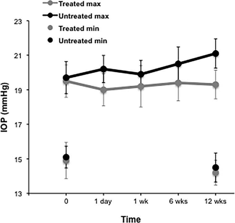
Aims: To evaluate temporal change in anterior chamber angle anatomy following argon laser peripheral iridoplasty (ALPI) in eyes with occludable angles postlaser peripheral iridotomy (LPI) compared with control eyes. Additionally, the effect on diurnal intraocular pressure (DIOP) fluctuation (maximum-minimum IOP) was investigated.
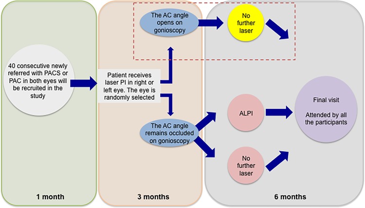
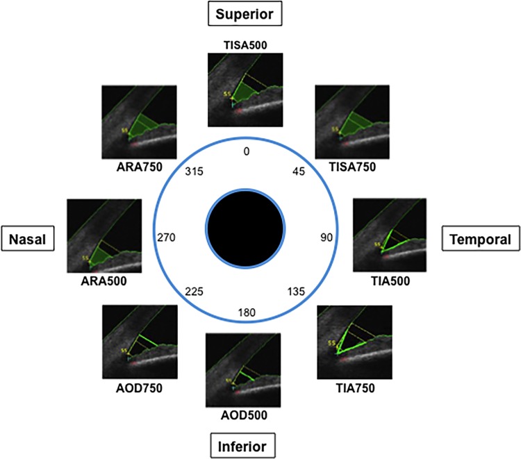
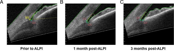
Methods: Twenty-two patients with bilateral primary angle closure/suspects with gonioscopically occludable anterior chamber angles following LPI were randomised to receive ALPI (n=11) or no further treatment (n=11). Angle opening distance (AOD), trabecular-iris angle, angle recess area and trabecular-iris space area were measured over eight sections with swept-source anterior segment optical coherence tomography and DIOP was measured pre-LPI and repeated at 3 months after ALPI (hourly measures).
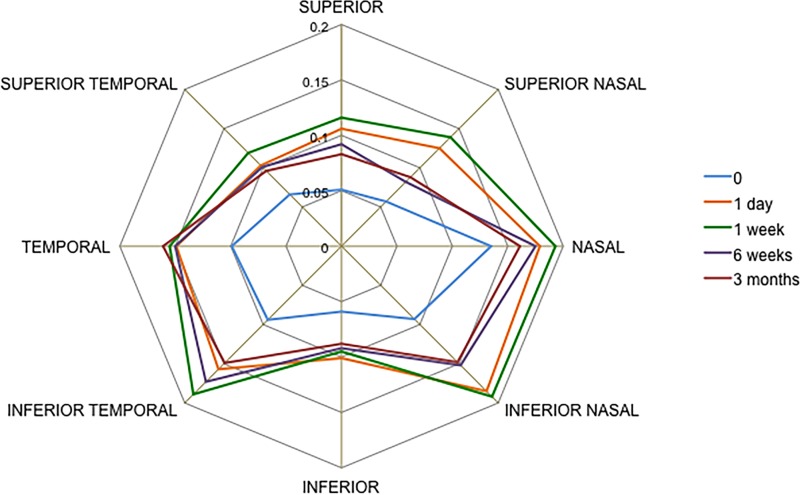
Results: All angle parameters increased following ALPI. This change was maintained for 3 months in seven of the eight sections (eg, inferotemporal AOD500 increased by 0.063 mm, p=0.004 at 1 day; 0.051 mm, p=0.029 at 1 week; 0.059 mm, p=0.006 at 6 weeks and 0.056 mm, p=0.011 at 3 months). The only exception was in the inferior sector (eg, AOD500 increased by 0.041 mm, p=0.025 at 1 day and by 0.029 mm, p=0.054 at 3 months). DIOP at 3 months was significantly reduced (5.04 mm Hg; ±1.61 mm Hg) compared with controls (6.61 mm Hg; ±1.63 mm Hg). Maximum IOP was significantly greater in the non-ALPI group (1.87 mm Hg, p=0.026).
Conclusions: ALPI widened all angle sections in eyes that remained occludable post-LPI. Changes were maintained for 3 months. ALPI decreased DIOP fluctuation in the treated eyes by lowering the maximum IOP value.
Keywords: Anterior chamber; Clinical Trial; Glaucoma; Imaging; Treatment Lasers.
Published by the BMJ Publishing Group Limited. For permission to use (where not already granted under a licence) please go to http://www.bmj.com/company/products-services/rights-and-licensing/.
Conflict of interest statement
Competing interests: None declared.
Figures

Similar articles
-
Optical coherence tomography-measured changes over time in anterior chamber angle and diurnal intraocular pressure after laser iridotomy: IMPACT study.Clin Exp Ophthalmol. 2018 Nov;46(8):895-902. doi: 10.1111/ceo.13303. Epub 2018 May 16. Clin Exp Ophthalmol. 2018. PMID: 29767420 Clinical Trial.
-
Effects of lens extraction versus laser peripheral iridotomy on anterior segment morphology in primary angle closure suspect.Graefes Arch Clin Exp Ophthalmol. 2019 Jul;257(7):1473-1480. doi: 10.1007/s00417-019-04353-8. Epub 2019 May 11. Graefes Arch Clin Exp Ophthalmol. 2019. PMID: 31079203 Clinical Trial.
-
Quantitative assessment of changes in anterior segment morphology after argon laser peripheral iridoplasty: findings from the EARL study group.Clin Exp Ophthalmol. 2019 Jan;47(1):33-40. doi: 10.1111/ceo.13374. Epub 2018 Sep 11. Clin Exp Ophthalmol. 2019. PMID: 30098125
-
Anterior Segment Optical Coherence Tomography Changes to the Anterior Chamber Angle in the Short-term following Laser Peripheral Iridoplasty.J Curr Glaucoma Pract. 2014 Jan-Apr;8(1):1-6. doi: 10.5005/jp-journals-10008-1152. Epub 2014 Jan 16. J Curr Glaucoma Pract. 2014. PMID: 26997799 Free PMC article. Review.
-
Changing patterns in treatment of angle closure glaucoma.Curr Opin Ophthalmol. 2018 Mar;29(2):130-134. doi: 10.1097/ICU.0000000000000453. Curr Opin Ophthalmol. 2018. PMID: 29194069 Review.
Cited by
-
Lasers in Glaucoma: an Overview.Int Ophthalmol. 2021 Mar;41(3):1111-1128. doi: 10.1007/s10792-020-01654-4. Epub 2020 Nov 19. Int Ophthalmol. 2021. PMID: 33211223 Review.
-
Lasers in glaucoma.Indian J Ophthalmol. 2018 Nov;66(11):1539-1553. doi: 10.4103/ijo.IJO_555_18. Indian J Ophthalmol. 2018. PMID: 30355858 Free PMC article. Review.
-
Laser peripheral iridoplasty for chronic angle closure.Cochrane Database Syst Rev. 2021 Mar 23;3(3):CD006746. doi: 10.1002/14651858.CD006746.pub4. Cochrane Database Syst Rev. 2021. PMID: 33755197 Free PMC article.
-
Efficacy and safety of laser peripheral iridoplasty with different energy levels and locations in the treatment of primary angle closure disease assessed by swept-source anterior segment optical coherence tomography.BMC Ophthalmol. 2023 Apr 11;23(1):149. doi: 10.1186/s12886-023-02899-0. BMC Ophthalmol. 2023. PMID: 37041488 Free PMC article. Clinical Trial.
-
Induction of significant intraocular pressure diurnal fluctuation in rats using a modified technique of microbead occlusion.Int J Ophthalmol. 2018 Jul 18;11(7):1114-1119. doi: 10.18240/ijo.2018.07.07. eCollection 2018. Int J Ophthalmol. 2018. PMID: 30046526 Free PMC article.
References
Publication types
MeSH terms
LinkOut - more resources
Full Text Sources
Other Literature Sources