. 2016 Dec 7;36(49):12425-12435.
doi: 10.1523/JNEUROSCI.2016-16.2016.

Vectored Intracerebral Immunization with the Anti-Tau Monoclonal Antibody PHF1 Markedly Reduces Tau Pathology in Mutant Tau Transgenic Mice

Affiliations

Vectored Intracerebral Immunization with the Anti-Tau Monoclonal Antibody PHF1 Markedly Reduces Tau Pathology in Mutant Tau Transgenic Mice

Wencheng Liu et al. J Neurosci. .

Erratum in

Abstract

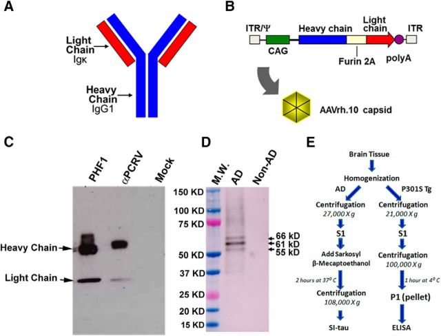
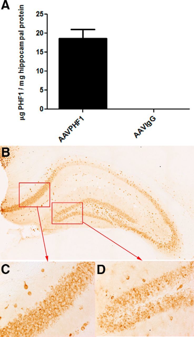
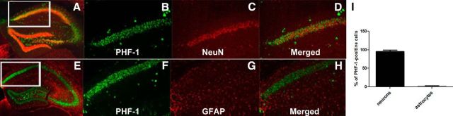
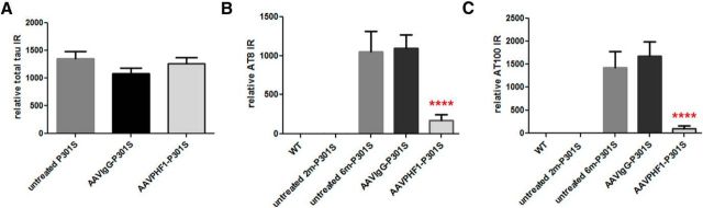
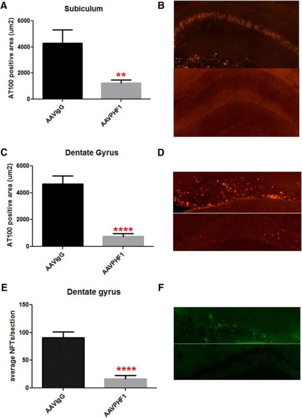
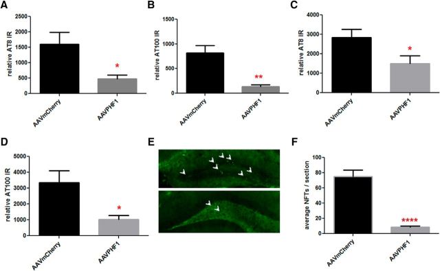
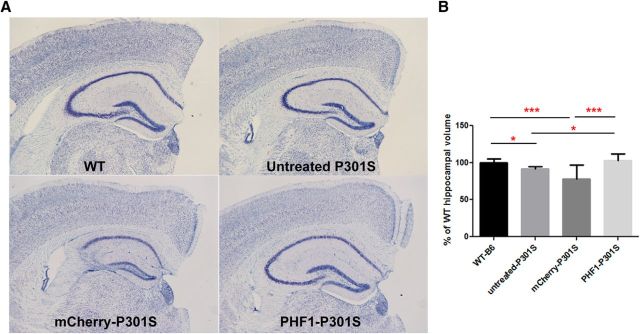
Passive immunization with anti-tau monoclonal antibodies has been shown by several laboratories to reduce age-dependent tau pathology and neurodegeneration in mutant tau transgenic mice. These studies have used repeated high weekly doses of various tau antibodies administered systemically for several months and have reported reduced tau pathology of ∼40-50% in various brain regions. Here we show that direct intrahippocampal administration of the adeno-associated virus (AAV)-vectored anti-phospho-tau antibody PHF1 to P301S tau transgenic mice results in high and durable antibody expression, primarily in neurons. Hippocampal antibody levels achieved after AAV delivery were ∼50-fold more than those reported following repeated systemic administration. In contrast to systemic passive immunization, we observed markedly reduced (≥80-90%) hippocampal insoluble pathological tau species and neurofibrillary tangles following a single dose of AAV-vectored PHF1 compared with mice treated with an AAV-IgG control vector. Moreover, the hippocampal atrophy observed in untreated P301S mice was fully rescued by treatment with the AAV-vectored PHF1 antibody. Vectored passive immunotherapy with an anti-tau monoclonal antibody may represent a viable therapeutic strategy for treating or preventing such tauopathies as frontotemporal dementia, progressive supranuclear palsy, or Alzheimer's disease.

Significance statement: We have used an adeno-associated viral (AAV) vector to deliver the genes encoding an anti-phospho-tau monoclonal antibody, PHF1, directly to the brain of mice that develop neurodegeneration due to a tau mutation that causes frontotemporal dementia (FTD). When administered systemically, PHF1 has been shown to modestly reduce tau pathology and neurodegeneration. Since such antibodies do not readily cross the blood-brain barrier, we used an AAV vector to deliver antibody directly to the hippocampus and observed much higher antibody levels and a much greater reduction in tau pathology. Using AAV vectors to deliver antibodies like PHF1 directly to brain may constitute a novel approach to treating various neurodegenerative disorders, such as FTD and Alzheimer's disease.

Keywords: AAV vector; Alzheimer's disease; PHF-tau; anti-tau antibody; passive immunization; tauopathy.


Figures

Figure 1.
Figure 1.
Diagram of the PHF1 vector, expression cassette, and in vitro expression of PHF1. A, Schematic of the full-length antibody, including light and heavy chains. B, Diagram of the AAVrh.10PHF1 genome. The full-length antibody expression cassette is flanked by the two inverted terminal repeats of AAV serotype 2 (ITR) and encapsidation signal (Ψ). The expression cassette comprises the following: the human cytomegalovirus (CMV) enhancer, the chicken β-actin promoter/splice donor and 5′ end of intron, the 3′ end of the rabbit β-globin intron and splice acceptor, the full-length PHF1 antibody sequence expressed in a single open reading frame (ORF) with an optimized Kozak sequence, and the polyadenylation/transcription stop signal from rabbit β-globin. The full-length antibody ORF includes the IgG1 leader peptide and variable and constant regions (heavy chain) in frame with the Igκ leader peptide, variable and constant regions (light chain), by inclusion of a furin cleavage recognition sequence upstream of a Tav 2A sequence (furin 2A). Expression of PHF1 was examined in the supernatant of 293T cells 48 h after transfection with pAAVPHF1. C, pAAVPHF1-transfected cells produce PHF1 full-length antibody secreted into the extracellular media. Western analysis lanes show supernatants from pAAVPHF1, pAAVαPCRV antibody control, and mock transfected cells. Arrows indicate antibody heavy and light chains. PHF1 and control antibodies were detected using a goat anti-mouse IgG antibody conjugated with HRP. D, PHF1 from the supernatant of transfected cells binds to pathogenic tau from an AD brain S1 fraction. Brain S1 fractions from non-AD and AD patients were separated by SDS PAGE (NuPAGE 4–12% Bis-Tris protein gels, Life Technologies) and assayed by Western blot using cell culture supernatants from pAAVPHF1-transfected 293T cells as a primary antibody and a goat anti-mouse IgG antibody conjugated to HRP as secondary antibody. Arrows indicated the three expected bands (55, 61, and 66 kDa proteins) for hyperphosphorylated/pathogenic Tau (p-Tau). A cortical S1 fraction from a non-AD subject was used as a negative control. M.W., Molecular weight standards. E, A schematic of biochemical procedures for obtaining human AD and P301S brain tissue fractions (details in Materials and Methods).
Figure 2.
Figure 2.
The presence of PHF1 in the hippocampus of P301S male mice treated with AAVrh.10PHF1 vector. A, PHF1, present in the soluble fraction of the hippocampus, is detected by binding to paired helical filamentous tau coated onto an ELISA plate. The antibody-antigen complex is visualized and quantified using HRP-labeled anti-mouse antibody. The complex and its TMB substrate were read at OD450 on a plate reader. PHF1 is not found in mice treated with the AAVrh.10IgG (anti-nicotine antibody) control vector. B, PHF1 antibody is detected in the hippocampus of P301S male mice treated with the AAVrh.10PHF1 vector by IHC using an anti-mouse IgG1 antibody. C, D, Higher-power images of the subiculum and DG regions (red boxes) of B. Note that PHF1 is a mouse IgG1.
Figure 3.
Figure 3.
PHF1 is expressed primarily in hippocampal neurons of P301S male mice treated with AAVrh.10PHF1 vector. A–H, PHF1 is primarily colocalized to NeuN-positive neurons (A–D) but not GFAP-positive astrocytes (D–H). A, A representative photomicrograph of double-staining IHC using an anti-mouse antibody (green) and NeuN antibody (red) and hippocampal sections from P301S mice treated with AAVrh.10PHF1. B, C, Higher-power images of PHF1 (B) and NeuN (C) expression from the boxed area of A. D, A merged image of B and C. E, A representative photomicrograph of double-staining IHC using an anti-mouse antibody (green) and GFAP antibody (red) and of hippocampal sections of P301S mice treated with AAVrh.10PHF1. F, G, Higher-power images of PHF1 (F) and GFAP (G) expression in the boxed area of E. H, A merged image of F and G. I, Quantitative analysis of the subiculum of AAVrh.10PHF1-treated P301S mice (n = 12) demonstrated that 95.5 ± 0.009% of PHF1-expressing cells are NeuN-positive neurons, whereas only 2.12 ± 0.32% of PHF1-expressing cells are GFAP-positive astrocytes.
Figure 4.
Figure 4.
Marked reduction of insoluble p-tau in the hippocampus of AAVrh.10PHF1-treated P301S male mice. S1 and P1 fractions from wild-type and various groups of P301S mice were prepared to measure total tau and p-tau, respectively. A, Total tau levels are not changed in AAVrh.10-treated P301S versus untreated male mice. B, P301S male mice do not display detectable AT8 immunoreactivity (IR) in the hippocampal insoluble fraction at 2 months of age, whereas a very high level of AT8 IR is present at 6 months of age. Insoluble p-tau levels in the hippocampus of 6-month-old P301S mice treated with the AAVrh.10IgG control vector are comparable to those of untreated P301S mice of the same age, but markedly reduced following treatment with AAVPHF1 (84%; p < 0.0001). C, Similar to the results found in the AT8 ELISA analyses in B, there is a significant reduction (93.8% reduction; p < 0.0001) in AT100 IR in the hippocampal insoluble fraction of AAVrh.10PHF1-treated P301S mice. Relative IR represents the total IR or p-tau IR detected in the indicated amount of S1 protein (in nanograms) from the brain homogenates of cases of AD (see Materials and Methods for details). **** p ≤ 0.0001.
Figure 5.
Figure 5.
Marked decrease of insoluble p-tau and NFTs in the subiculum and DG of AAVrh.10PHF1-treated P301S male mice. A, C, The area of AT100-positive immunoreactivity (IR) is significantly reduced in the subiculum (A, ∼72% reduction, p = 0.0025) and DG (C, ∼84% reduction, p < 0.0001) of AAVrh.10PHF1-treated P301S male mice. B, D, Representative photomicrographs for A (B) and C (D). Top, AT100 immunofluorescence in the subiculum and DG, respectively, of P301S mice treated with the AAVrh.10IgG control vector. Bottom, AT100 IR of P301S mice treated with AAV rh.10PHF1 vector. E, Thioflavin-S staining was used to visualize and quantify NFTs, comprising abnormally misfolded and hyperphosphorylated tau in the DG of AAVrh.10IgG control and AAVrh10PHF1-treated P301S mice. Quantitative analysis demonstrates that the number of NFTs is significantly reduced in the DG of AAVrh.10PHF1-treated mice compared with control mice (∼83% reduction, p < 0.0001). F, A representative image of E. Top, Thioflavin-S staining of the DG from mice treated with the control vector. Bottom, Staining of the DG from AAVrh.10PHF1-treated P301S mice. All quantitative analyses were performed blind to treatment (A, C, and E). ** 0.001 < p ≤ 0.01; **** p ≤ 0.0001.
Figure 6.
Figure 6.
Marked reduction in insoluble p-tau and NFTs in the DG of AAVrh.10-treated P301S female mice. A, B, The levels of AT8 IR (A; ∼70% reduction; p = 0.05) and AT100 IR (B; 84% reduction; p = 0.003) are markedly reduced in the hippocampal insoluble fraction of AAVrh.10PHF1-treated P301S female mice (n = 7) compared with the AAVrh.10mCherry control mice (n = 13). C, D, We also observed a reduction of AT8 immunoreactivity (IR; C: ∼47%; p = 0.05) and AT100 IR (D: ∼70%; p = 0.04) in the cortical insoluble fraction from AAVPHF1-treated P301S compared with those of mCherry-treated P301S mice. E, Thioflavin-S staining was used to quantify the number of NFTs. Top, A representative photomicrograph of NFTs in the DG of P301S female mice treated with the AAVrh.10mCherry control vector. Bottom, A photomicrograph of the DG from AAVrh.10PHF1 vector-treated mice. F, Quantitative analysis performed blind to treatment demonstrates that the number of NFTs is significantly reduced in the DG of AAVrh.10PHF1-treated P301S female mice (∼88% reduction; n = 13; p < 0.0001). * 0.01 < p ≤ 0.05; ** 0.001 < p ≤ 0.01; **** p ≤ 0.0001.
Figure 7.
Figure 7.
Hippocampal atrophy in P301S mice is rescued by treatment with AAVrh.10PHF1. Hippocampal volumes are not changed between AAVrh.10PHF1-treated P301S female mice and wild-type mice, indicating that there is no obvious neurotoxicity following AAVrh.10PHF1 treatment. Hippocampal volumes were measured using three coronal brain sections from each animal at the following level relative to bregma: −1.7, −1.94, and −2.30. A, A representative Nissl-stained hippocampal section from a wild-type female mouse as well as from untreated, AAVrh.10mCherry-treated, and AAVrh.10PHF1-treated P301S female mice. B, Statistical analysis demonstrates that there is a decrease in hippocampal volume (∼8% reduction; p = 0.01) in P301S female mice at 6 months of age compared with that of aged-matched wild-type female control mice. A more significant reduction in hippocampal volume (∼21%) was observed in AAVrh.10mCherry-treated P301S mice (p = 0.0006) compared with that of wild-type mice. No significant difference in hippocampal volume was observed between AAVrh.10PHF1-treated and wild-type female mice. Importantly, compared with the hippocampal volume of untreated P301S female mice, treatment with the AAVrh.10PHF1 vector fully rescued this loss of hippocampal volume. Hippocampal volumes were measured using NIS software (Nikon). * 0.01 < p ≤ 0.05; *** 0.0001 < p ≤ 0.001.

References

    1. Ahmed Z, Cooper J, Murray TK, Garn K, McNaughton E, Clark H, Parhizkar S, Ward MA, Cavallini A, Jackson S, Bose S, Clavaguera F, Tolnay M, Lavenir I, Goedert M, Hutton ML, O'Neill MJ. A novel in vivo model of tau propagation with rapid and progressive neurofibrillary tangle pathology: the pattern of spread is determined by connectivity, not proximity. Acta Neuropathol. 2014;127:667–683. doi: 10.1007/s00401-014-1254-6. - DOI - PMC - PubMed
    1. Allen B, Ingram E, Takao M, Smith MJ, Jakes R, Virdee K, Yoshida H, Holzer M, Craxton M, Emson PC, Atzori C, Migheli A, Crowther RA, Ghetti B, Spillantini MG, Goedert M. Abundant tau filaments and nonapoptotic neurodegeneration in transgenic mice expressing human P301S tau protein. J Neurosci. 2002;22:9340–9351. - PMC - PubMed
    1. Arriagada PV, Growdon JH, Hedley-Whyte ET, Hyman BT. Neurofibrillary tangles but not senile plaques parallel duration and severity of Alzheimer's disease. Neurology. 1992;42:631–639. doi: 10.1212/WNL.42.3.631. - DOI - PubMed
    1. Atwal JK, Chen Y, Chiu C, Mortensen DL, Meilandt WJ, Liu Y, Heise CE, Hoyte K, Luk W, Lu Y, Peng K, Wu P, Rouge L, Zhang Y, Lazarus RA, Scearce-Levie K, Wang W, Wu Y, Tessier-Lavigne M, Watts RJ. A therapeutic antibody targeting BACE1 inhibits amyloid-beta production in vivo. Sci Transl Med. 2011;3:84ra43. doi: 10.1126/scitranslmed.3002254. - DOI - PubMed
    1. Baek RC, Broekman ML, Leroy SG, Tierney LA, Sandberg MA, D'Azzo A, et al. AAV-mediated gene delivery in adult GM1-gangliosidase mice corrects lysosomal storage in CNS and improves survival. PloS One. 2010 doi: 10.1371/journal.pone.0013468. 5.e13468. - DOI - PMC - PubMed

MeSH terms